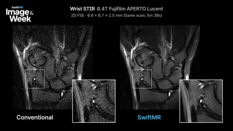
Improving Wrist STIR on a 0.4T Open MRI
A low-field wrist STIR MRI had a signal problem. The solution didn't require a higher-field scanner.
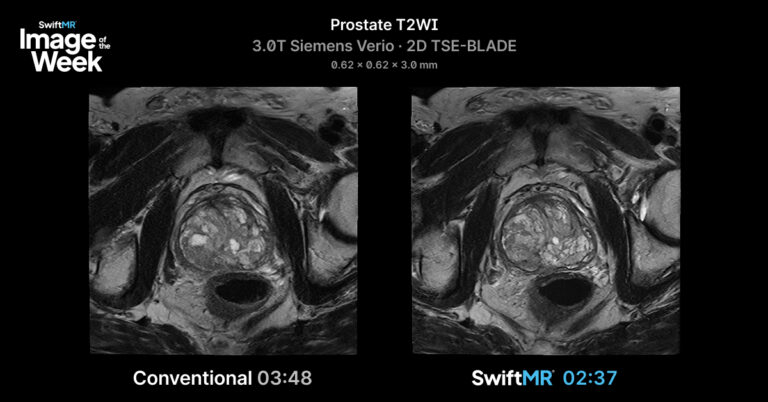
Prostate T2-Weighted Imaging in 31% Less Time
On a 3T Siemens Verio, SwiftMR cut a prostate T2 scan from 3:48 to 2:37 – 31% faster, but at the same resolution. Less time on the table means less chance...
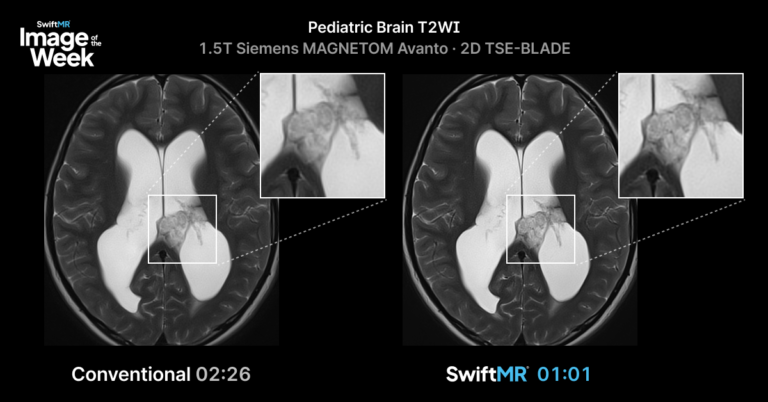
Pediatric Brain T2WI MRI: Evaluating a 58% Shorter Acquisition with SwiftMR
In pediatric MRI, every minute on the table matters.
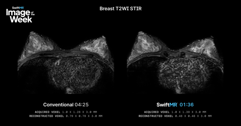
Breast T2WI STIR MRI: Evaluating Accelerated Acquisition with SwiftMR
Breast MRI requires image quality, patient cooperation, and consistency across sequences. In prone positioning, longer acquisitions can increase the opportunity...
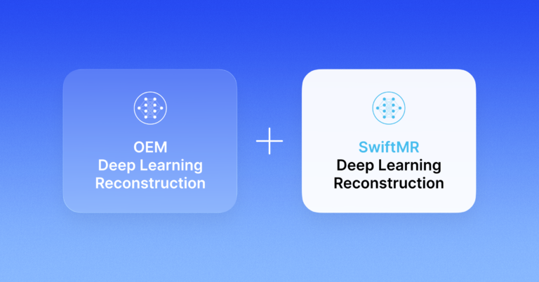
How SwiftMR Extends OEM DL Beyond Current Limitations
Discover how OEM DL reconstruction and SwiftMR work together to deliver broader MRI image quality and acceleration benefits.
SwiftMR for Hitachi Open MRI Systems: AIRIS 0.3T & OASIS 1.2T
SwiftMR helps Hitachi AIRIS 0.3T and OASIS 1.2T open MRI systems reduce scan times, improve image quality, and increase scanner capacity without new hardware....
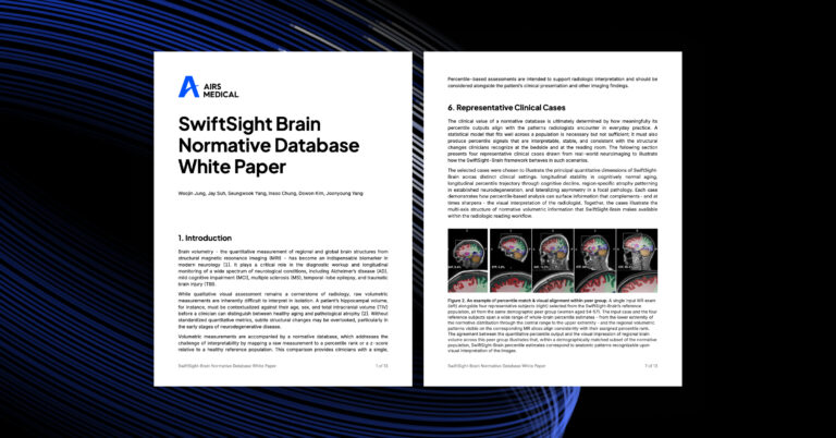
SwiftSight-Brain Normative Database for Quantitative Brain MRI
Now updated with the world’s largest normative database in quantitative brain MRI, built from 32,615 subjects across vendors, field strengths, and international...
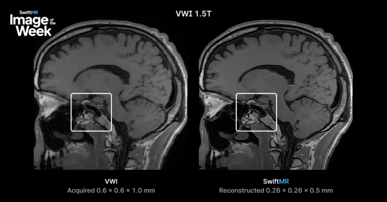
1.5T Vessel Wall Imaging: Evaluating Reconstructed Detail
Intracranial vessel wall imaging is technically demanding. The vessel wall is small, adjacent blood signal must be suppressed, and image quality depends...
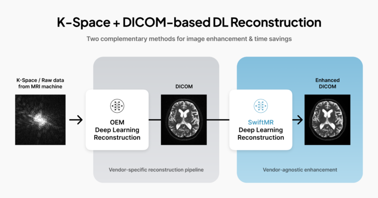
7 Ways SwiftMR Improves OEM DL Reconstruction for Radiology Practices
OEM deep learning reconstruction has changed what MRI teams can expect from accelerated imaging.
By improving reconstruction within the scanner pipeline,...
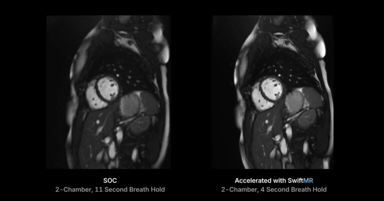
AI-Accelerated Cardiac MRI: 69% Faster Short-Axis Cine With SwiftMR
Cardiac MRI has never been more clinically important — or more operationally demanding.
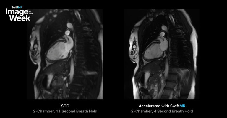
Cardiac Cine MRI in 4 Heartbeats: Reducing Breath-Hold Burden in CMR
SwiftMR reconstruction reduces 2-chamber cine acquisition from 11 heartbeats to 4, cutting breath-hold time by more than 60% while maintaining diagnostic...
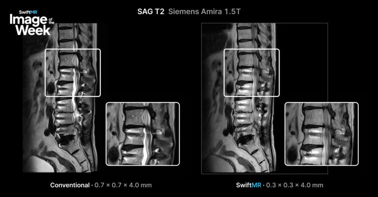
Metal Artifact Reduction MRI: Recovering Signal with SwiftMR
Metal Hardware MRI: Same Protocol. Recovering Signal Where It Counts
Metal hardware is one of the biggest image quality challenges in MRI, and one of the...

Beyond Faster Scans: Where We're Taking MRI Next
Following the TA Associates growth investment, AIRS Medical CEO Jason Park shares why the future of MRI is measurable insight, not just faster scans.
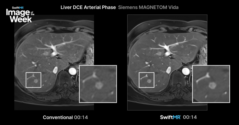
Liver DCE MRI Evaluating Vessel Delineation and Lesion Conspicuity
Liver DCE MRI — Same Scan. More to See.
Arterial phase imaging requires precise timing and sufficient image quality to evaluate enhancement patterns, hepatic...
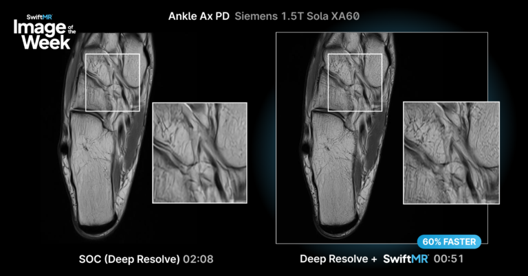
Deep Resolve and SwiftMR: Accelerated Ankle MRI to 51 Seconds
Ankle Ax PD
Musculoskeletal MRI often requires balancing image quality with efficiency. This example compares standard deep resolve AI and accelerated...