Clinical Evidence
Peer-reviewed results and before-and-after comparisons for SwiftMR® and SwiftSight-Brain.
Denoising
Improve SNR with well-preserved tissue contrast
Super-resolution
Sharper lesion and structural conspicuity
Acceleration
Shorter scan time, less motion artifact
Artifact reduction
Cleaner truncation and susceptibility artifacts
MS lesion tracking
New, enlarging, and stable lesions across exams
Diagnostic Performance
Peer-reviewed studies that measured diagnostic performance in the same patients with and without SwiftMR.
47% faster scans, with higher sensitivity for foraminal stenosis
Long scans are hard on patients with spinal disease and invite motion artifacts. Each patient had a conventional scan and one 47 to 48% shorter with SwiftMR on the same day, read by four radiologists.
Sensitivity for neural foraminal stenosisSensitivity for neural foraminal stenosis: Conventional, 65%. With SwiftMR, 73%. Difference 7 points (95% CI 2–13), p = .005.
Difference 7 points (95% CI 2–13), p = .005
More accurate glioma grading from DCE perfusion maps
DCE-MRI trades signal-to-noise for temporal resolution, which adds variability to perfusion measurements. With SwiftMR, mean extracellular volume (Ve) separated low- from high-grade diffuse glioma more accurately.
AUROC for WHO grade, mean VeAUROC for WHO grade, mean extracellular volume: Standard DCE, 0.83 (95% CI 0.76–0.90). With SwiftMR, 0.88 (95% CI 0.83–0.94). p = .02.
Lee et al. Scientific Reports, 2024
More accurate TLE diagnosis on thin‑slice MRI
Thin slices show the hippocampus in more detail but add noise. With SwiftMR, two neuroradiologists diagnosed TLE more accurately from the same 1.5 mm coronal T2-weighted images.
Pooled diagnostic accuracy, 1.5 mm imagesPooled diagnostic accuracy, 1.5 mm images: Without SwiftMR, 73.1% (95% CI 65.5–79.5). With SwiftMR, 80.8% (95% CI 74.0–86.1). p = .010, pooled across both readers.
p = .010, pooled across both readers
Suh et al. Korean Journal of Radiology, 2024
Intracranial vessel wall imaging
Three times the sensitivity for culprit plaques
Limited signal-to-noise makes plaque features hard to see, especially in small distal arteries. Using intraplaque hemorrhage, sensitivity for culprit plaques rose from 23.5% to 70.6% with SwiftMR, and specificity held at 94.3%.
AUROC for culprit plaque, intraplaque hemorrhageAUROC for culprit plaque, intraplaque hemorrhage: Conventional, 0.59 (95% CI 0.48–0.70). With SwiftMR, 0.82 (95% CI 0.71–0.94). p < .001, DeLong test.
Seo et al. Scientific Reports, 2024
Values as published, with 95% confidence intervals (whiskers) where the study reported them. AUROC bars start at 0.5, the value for a test no better than chance. Each study compared images of the same patients with and without SwiftMR.
Improved SNR with well-preserved tissue contrast, revealing structures that noise obscures, without changing your scan protocol.
Deep learning improves quality of intracranial vessel wall MRI for better characterization of potentially culprit plaques
SwiftMR provided significant improvement in SNR and spatial resolution in vessel wall imaging, leading to detection of vessel wall lesions and intraplaque hemorrhage compared to conventional image.
Seo et al.. Scientific Reports. Aug 2024.
Improved lesion and structural conspicuity, enabling thin-slice imaging that would otherwise be limited by SNR constraints.
Improving diagnostic performance of MRI for temporal lobe epilepsy with deep learning-based image reconstruction in patients with suspected focal epilepsy
SwiftMR resulted in significantly improved resolution and structural conspicuity in hippocampal imaging compared to routine imaging. Hippocampal striation blurring was suspected on routine 3mm MRI, but became more obvious on 1.5mm MRI + SwiftMR.
Suh et al.. Korean Journal of Radiology. 2024;25(4):374-383.
Shorter scan time means less patient movement, minimizing chances of motion artifacts, directly improving image quality as a secondary benefit.
Deep learning-based reconstruction for acceleration of lumbar spine MRI: a prospective comparison with standard MRI
Prospective comparison showing 32.3% average acquisition time saving versus standard protocol, with no degradation in image quality or diagnostic performance.
Yoo et al.. Eur Radiol. 2023;33(12):8656-8668.
Highly accelerated knee magnetic resonance imaging using deep neural network (DNN)-based reconstruction: prospective, multi-reader, multi-vendor study
Prospective, multi-reader, multi-vendor study demonstrating average scan time reduction of 41% using SwiftMR, without degrading image quality or diagnostic performance across readers or scanner platforms.
Lee et al.. Sci Rep. 2023;13:17264. PMID: 37828048.
Artifact Reduction
Improve truncation and susceptibility artifacts, producing cleaner images from the same acquisition without reacquisition or protocol changes.
Susceptibility artifacts reduction
Geometric distortion improvement
Brain DWI b1000 · 2D EPI Diffusion · Same acquisition3.0T Siemens MAGNETOM Skyra
1:30 — Without SwiftMR · 1.5×1.9×3.0 mm
1:30 — With SwiftMR · 1.5×1.9×3.0 mm
Truncation artifact improvement
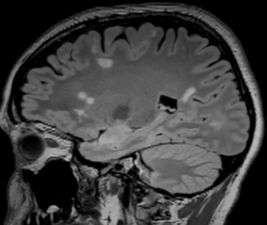
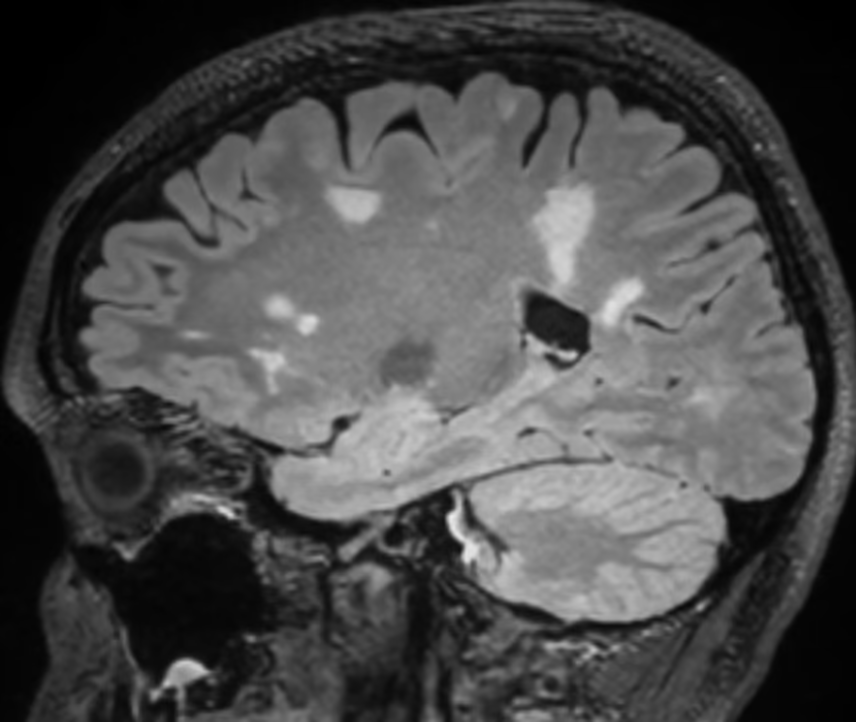
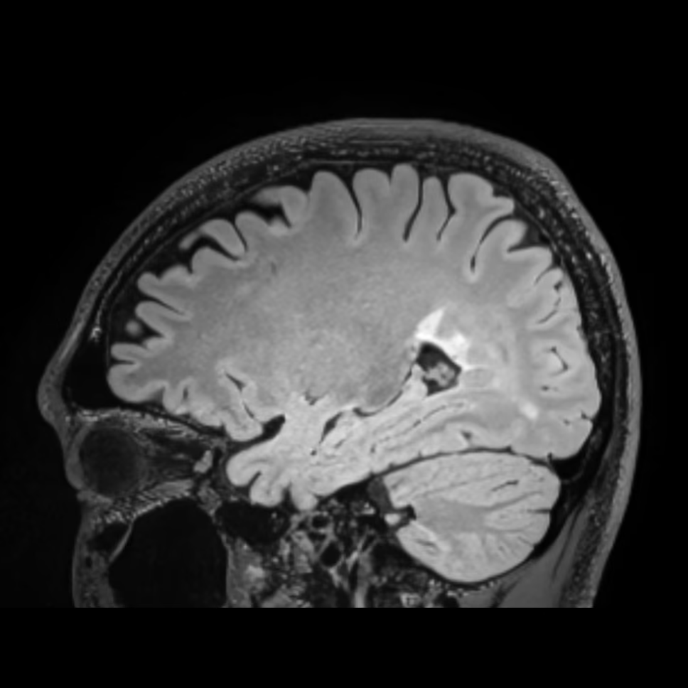
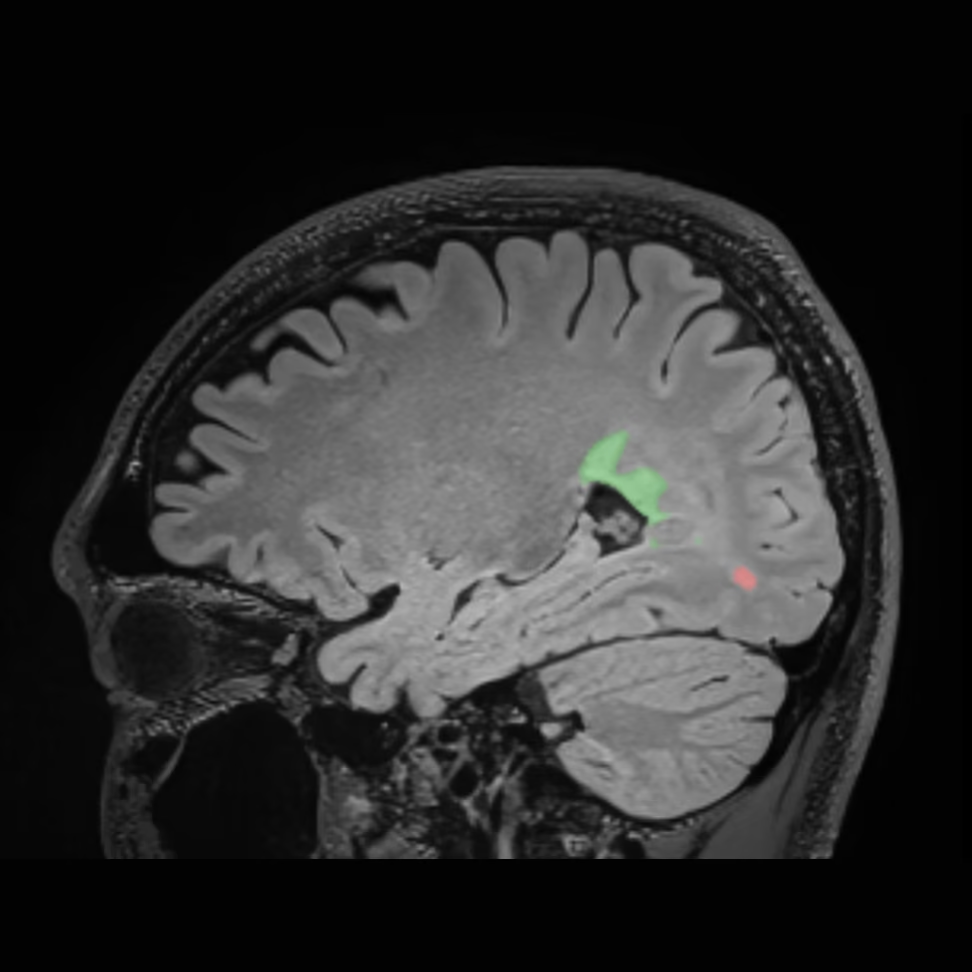
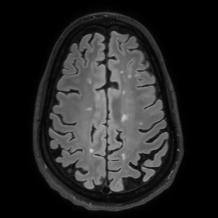
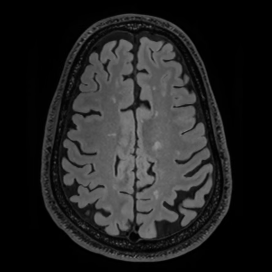
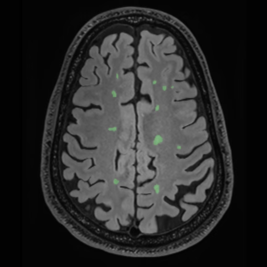
MS Lesion Tracking
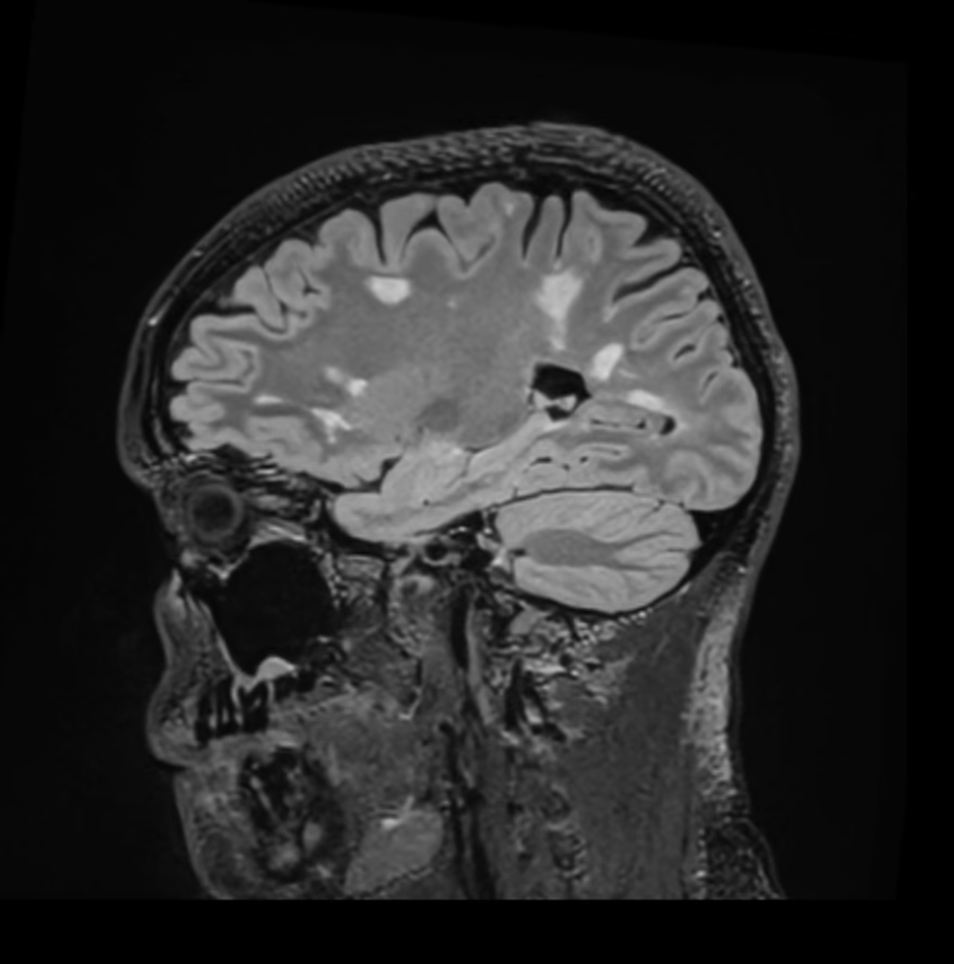
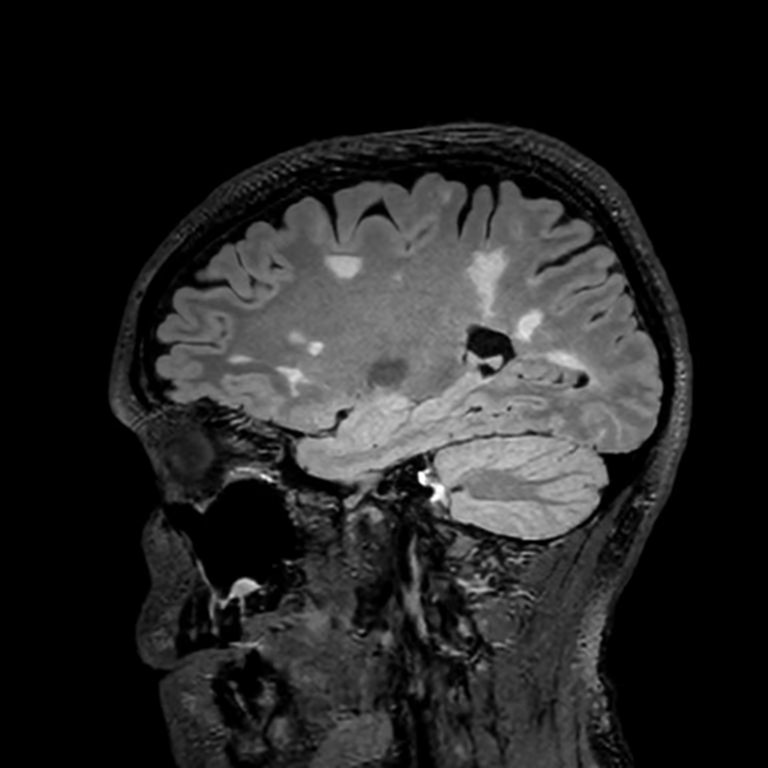
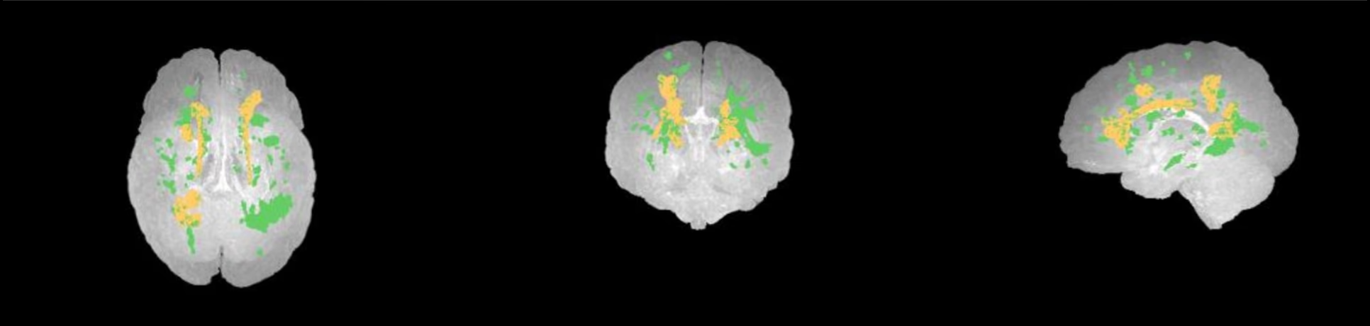
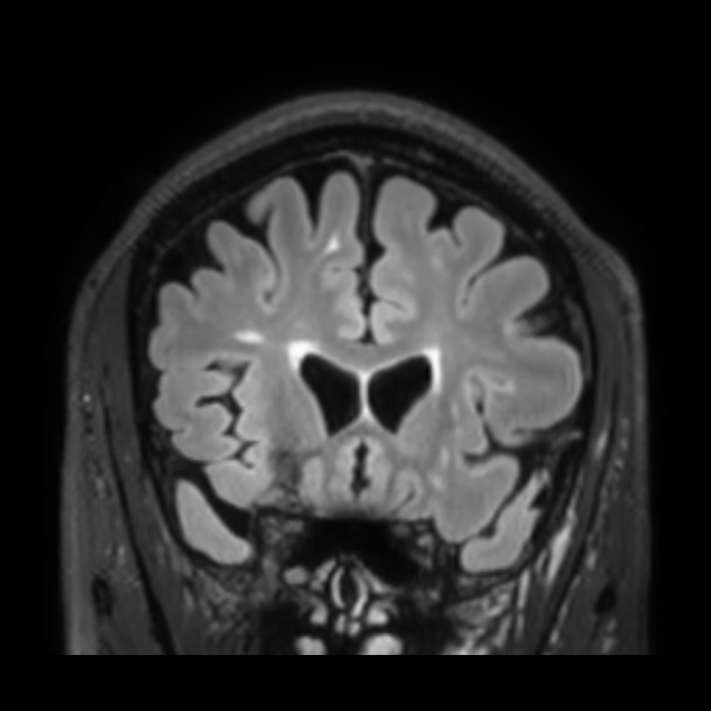
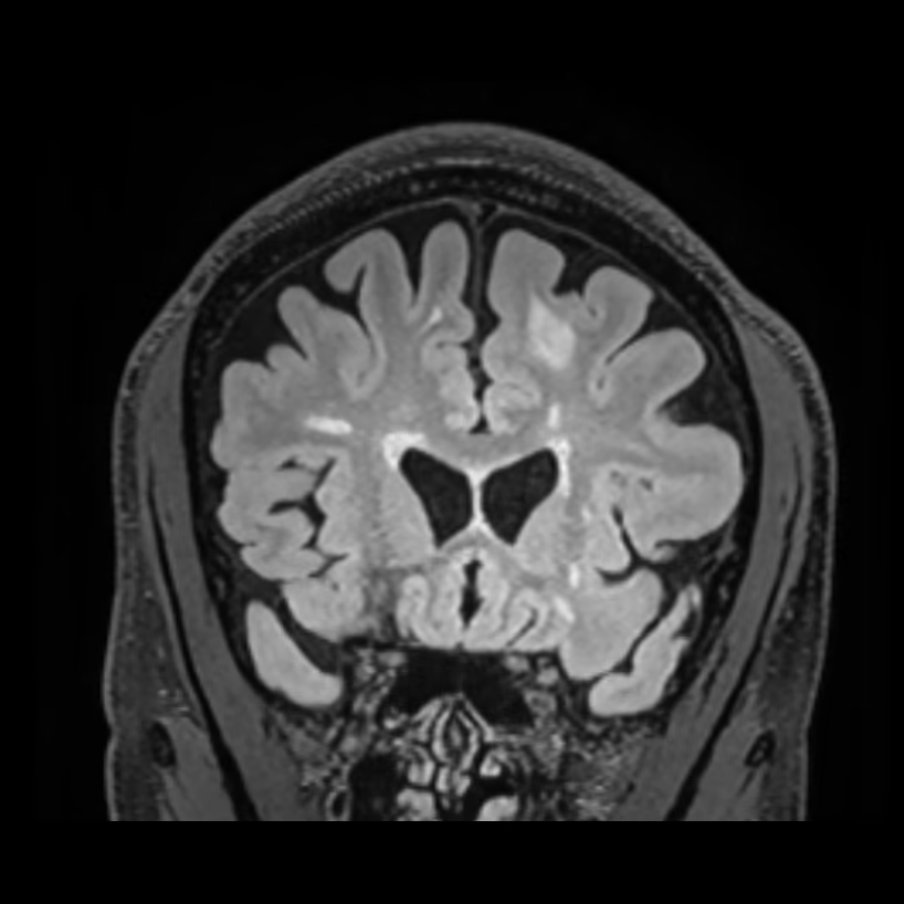
Longitudinal MS tracking compares exams taken months or years apart, often on different scanners. A change in vendor, field strength, or protocol can alter noise and resolution, so automated measurements can shift even when the patient has not changed.
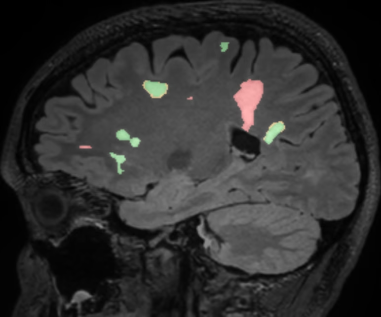
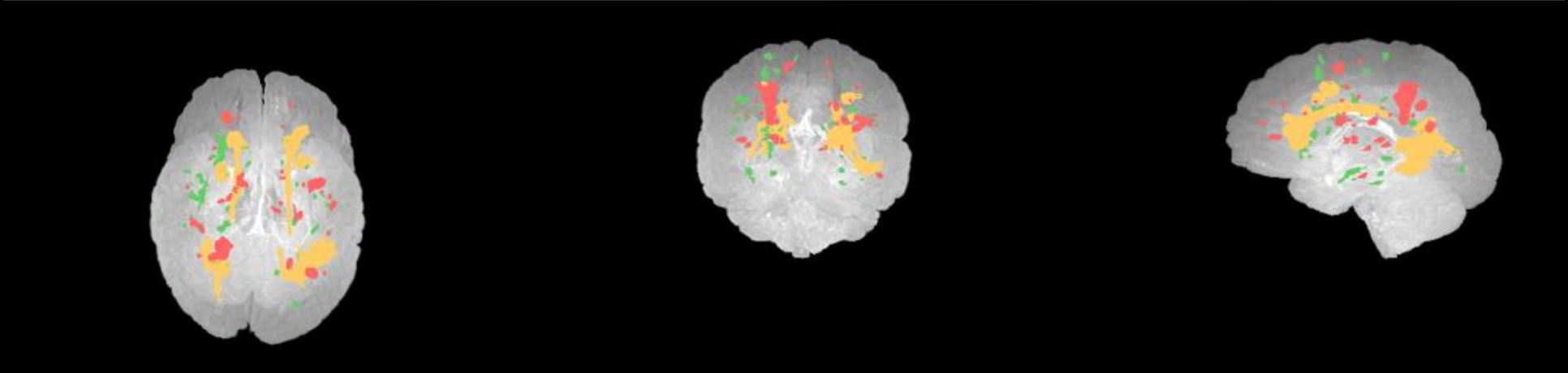
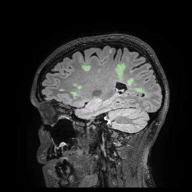
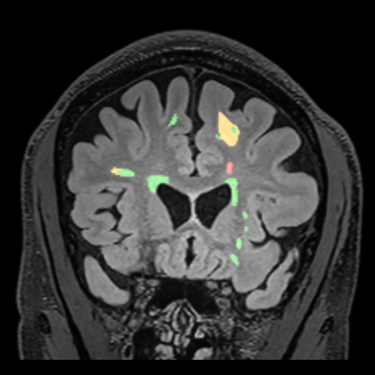
SwiftSight-Brain minimizes cross-scanner variation by normalizing and improving image quality before it compares exams, then classifies each lesion as new, enlarging, or stable.
Below are three examples of real-world variability between exams: a different scanner model, a different field strength, and a different vendor. In each, SwiftSight-Brain tracks the lesions consistently despite the change.
Cross-scanner variation in brain volumetry
Largest difference in hippocampal volume for one subject scanned on three scanners.
Across scanner models
Retrospective case · Female patient, age 31–39Siemens MAGNETOM Skyra 3T and Vida 3T
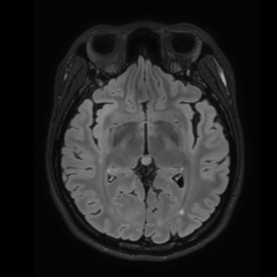
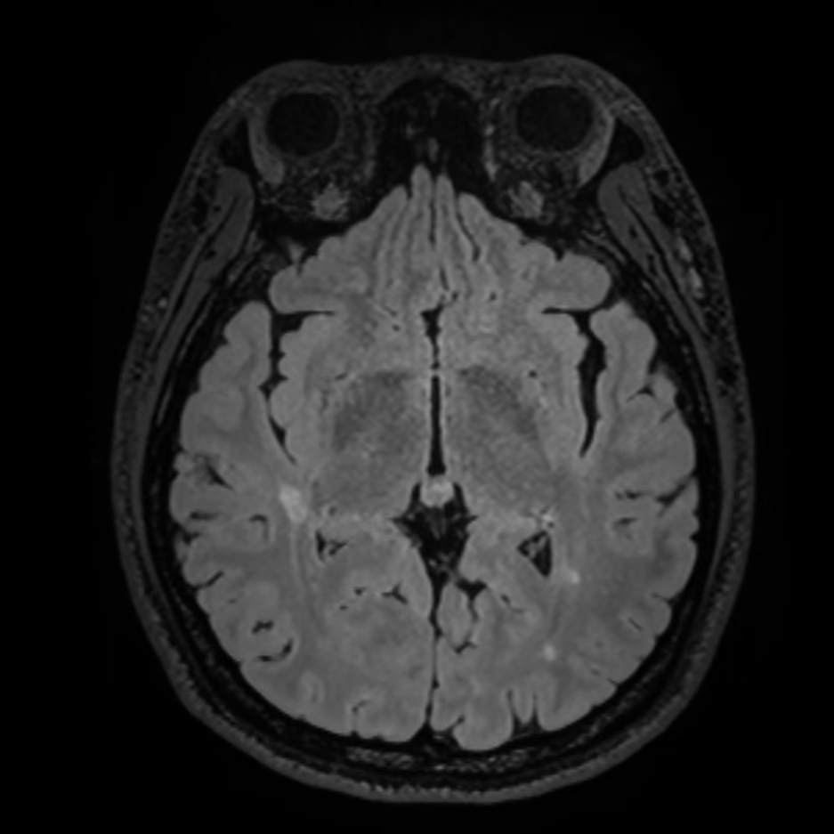
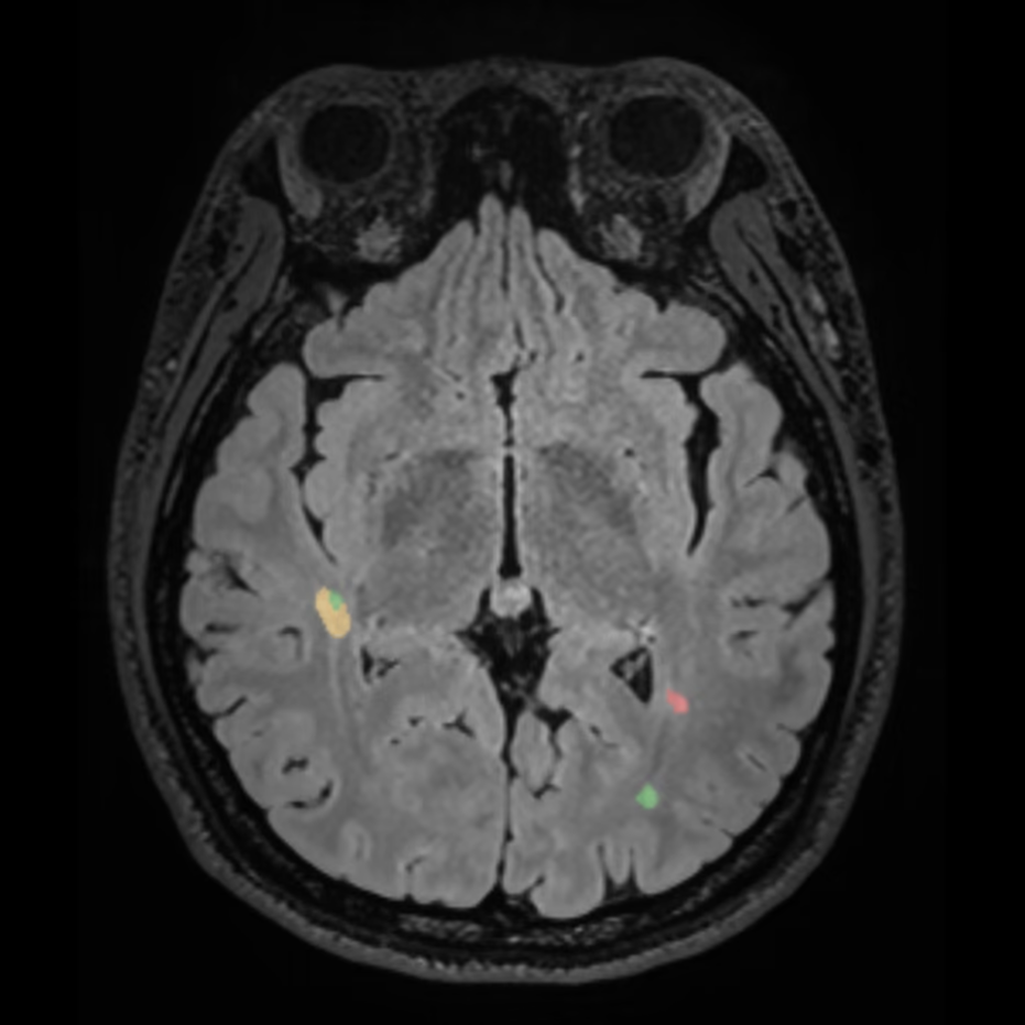
This patient's baseline at age 31 was acquired on a Siemens Skyra 3T, and the follow-up eight years later on a Vida 3T. SwiftSight-Brain found 32 lesions in the follow-up that were absent from the baseline, most of them in the deep white matter. Seven lesions that were already present, all periventricular, had grown, and their combined volume had more than tripled (+225.9%).
| Lesion type | Volume (ml) | Count | Change |
| New lesions | 2.85 | +32 | – |
| Enlarging lesions | 6.75 | +7 | +225.9% |
| Region | Volume (ml) | New | Enlarging |
| Periventricular | 11.71 | +9 | +7 |
| Deep white matter | 0.52 | +20 | 0 |
| Juxtacortical | 0.68 | +3 | 0 |
| Infratentorial | 0.0 | 0 | 0 |
| Whole brain | 12.91 | +32 | +7 |
Across field strengths and scanner models
Retrospective case · Female patient, age 38–39Siemens MAGNETOM Sola 1.5T and Vida 3T
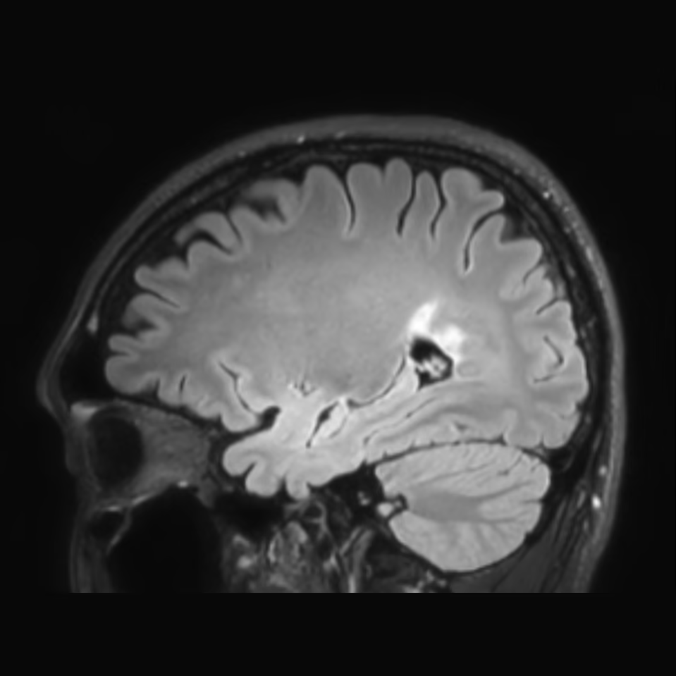
This patient's annual follow-ups were acquired on two Siemens scanners that differ in both model and field strength. Most lesions remained stable, and the largest lesion, in the parietal lobe, grew slightly. SwiftSight-Brainclassified that lesion as enlarging. The conventional solution classified it as stable.
| Lesion type | Volume (ml) | Count | Change |
| New lesions | 0 | 0 | – |
| Enlarging lesions | 1.36 | +5 | +12.2% |
| Region | Volume (ml) | New | Enlarging |
| Periventricular | 11.71 | 0 | +5 |
| Deep white matter | 0.52 | 0 | 0 |
| Juxtacortical | 0.68 | 0 | 0 |
| Infratentorial | 0.0 | 0 | 0 |
| Whole brain | 12.91 | 0 | +5 |
Across vendors
Retrospective cases · Philips 3T baseline, Siemens 3T follow-up
Vendor differences in noise and resolution can degrade segmentation accuracy. In four more patients with a Philips baseline and a Siemens follow-up, 11 months to more than three years apart, SwiftSight-Brain lesion quantification held steady despite the change of vendor.
FemaleAge 42–43Feb 2019 – Nov 2020Coronal view
FemaleAge 24–27Jan 2020 – May 2023Axial view
MaleAge 26–27Jul 2020 – Jun 2021Sagittal view
MaleAge 47–48Feb 2020 – Jan 2021Axial view
Request a Clinical Review
Our clinical team can provide in-depth clinical and technical presentations on SwiftMR.
Select publications
This white paper outlines AIRS Medical's information security practices for SwiftMR, and covers data workflow, encryption, disaster recovery, AI governance, and certifications. Cloud-based SwiftMR removes sensitive PHI from the DICOM tags as part of the de-identification process before transmitting the data to the SwiftMR Cloud Server.
This white paper quantifies the measurement consistency of SwiftSight-Brain, benchmarked against an established open-source reference, along four axes of variation a deployed volumetry tool encounters in practice: repeated acquisition on a fixed scanner, a change in magnetic field strength (1.5T versus 3T), a change of scanner hardware, and a change in in-plane spatial resolution.
In 86 patients scanned at 3T, accelerated T2-weighted liver imaging reconstructed with SwiftMR in a single breath-hold had higher SNR and sharpness than the routine two-breath-hold protocol, with lower spatial mismatch scores.
This white paper details the reference population and statistical methodology behind SwiftSight-Brain volumetric analysis, including a novel shifted-softplus normative model for age- and sex-adjusted assessment. The database draws on 3D T1-weighted MRI across major scanner vendors and field strengths to deliver stable, clinically interpretable percentile and z-score comparisons.
Across 173 patients, SwiftMR-reconstructed DCE series improved image noise, capsule sharpness, and lesion conspicuity for both readers, while PI-RADS diagnostic performance was preserved.
This white paper outlines AIRS Medical's information security practices for SwiftSight-Brain, covering the workflow surrounding data security, key security measures for secure data transmission and access control, and information security certifications and attestations. The security measures are designed to comply with applicable regulatory frameworks, including HIPAA, GDPR, and SOC 2 Type II requirements.
A two-institution prospective study of 70 patients found SwiftMR-enhanced accelerated lumbar spine MRI diagnostically interchangeable with conventional imaging at approximately 80 to 86% shorter acquisition time, with higher SNR and CNR, and automated reporting agreeing closely with radiologist consensus.
This study evaluated SwiftMR for improving the image quality and diagnostic performance of 3D contrast-enhanced T1-weighted black blood MRI for brain metastases. Image quality and noise improved, and sensitivity increased, particularly for lesions smaller than 5 mm, while specificity remained comparable.
In 50 patients, accelerated cervical spine MRI reconstructed with SwiftMR reduced acquisition time by 47 to 48%, had higher or similar image quality compared with the conventional protocol, and raised average sensitivity for neural foraminal stenosis from 65% to 73%.
In 306 patients with diffuse glioma, SwiftMR-enhanced DCE-MRI had higher SNR and CNR, more reliable arterial input function measurements, and improved differentiation of WHO grades based on the extravascular-extracellular volume fraction (AUROC 0.88 vs. 0.83).
This white paper compares accelerated MR images processed with SwiftMR to the standard of care in a multi-center, multi-reader study of 184 exams across neuro, musculoskeletal, and body imaging. Eighteen board-certified radiologists from six subspecialties scored the SwiftMR-processed images higher than the standard of care in 74.7% of cases.
This white paper models the economic value of SwiftMR for a hypothetical U.S. outpatient imaging center with one MRI scanner. In the base case, increased scan volume produced $271,790 in additional revenue over one year, an ROI of 453%, and a payback period of 3 months.
This study applied SwiftMR to 4D time-resolved contrast-enhanced MRA in 520 patients evaluated for ischemic stroke or intracranial vessel stenosis. Processed images showed better image quality, sharpness, vascular conspicuity, and SNR, with a better confidence level and shorter decision time for large vessel occlusion.
This paper introduces an all-in-one deep learning framework for MR image reconstruction, in which a single DICOM-based model enhances image quality across multiple aspects of k-space sampling. The algorithm serves as the core of SwiftMR and is compatible with various vendors, pulse sequences, scan parameters, and anatomical regions.
White paper by Geunu Jeong, MD, Head of SwiftMR Research at AIRS Medical, on protocol-setting strategies for scan time reduction, spatial resolution improvement, and SNR improvement. Its guiding principle: adjust scan parameters by willingly sacrificing either SNR or spatial resolution, and SwiftMR will compensate for the loss.
Peer-reviewed review paper exploring existing literature on the diverse applications of deep learning reconstruction (DLR) techniques for fast MR neuroimaging.
Retrospective study of 117 patients evaluating 1.5-mm coronal T2-weighted MRI with deep learning-based reconstruction (SwiftMR) for temporal lobe epilepsy. Pooled diagnostic accuracy on the same 1.5-mm images was 80.8% with SwiftMR versus 73.1% without it.
Review article on radiology as a major cost driver and the productivity gains and cost savings AI could bring, using Singapore healthcare as an example. It cites SwiftMR as an AI reconstruction tool that shortens MRI scan times without additional hardware.
This study evaluated the performance of SwiftMR in accelerating knee MRI without degrading image quality or diagnostic performance.
Prospective study of 50 patients with degenerative lumbar spine disease at 1.5T. The accelerated protocol with deep learning reconstruction reduced average acquisition time by 32.3% without degrading image quality or readers’ diagnostic performance.
For a full list of publications, contact bd@airsmed.com.
Ready to see SwiftMR® at your institution?
Our clinical team can provide in-depth presentations on SwiftMR — including protocol-specific performance data for your scanner and field strength.