3:00 → 1:30: Accelerating Non-Contrast 3D Aortic Arch MRA with Deep Learning Reconstruction
Non-contrast 3D MRA of the aortic arch can add valuable anatomical information to a cardiac MRI examination without the use of a gadolinium-based contrast agent. But acquiring that information efficiently is not always easy.
High-resolution 3D aortic arch imaging relies on respiratory and ECG gating, making the acquisition particularly sensitive to motion. It can also come later in a CMR examination, when the patient has already spent significant time in the scanner and tolerance may be declining.
The longer the acquisition, the greater the opportunity for motion, inconsistent gating, and image degradation that can potentially lead to repeat imaging.
In this case, SwiftMR® deep learning reconstruction was combined with parallel imaging, in-plane super-resolution, and through-plane super-resolution to substantially shorten the starting acquisition time while maintaining high-resolution visualization of the aortic arch.
A 50% reduction in starting acquisition time
This case was acquired on a Siemens Sola 1.5T, combining parallel imaging, in-plane super-resolution, through-plane super-resolution, and SwiftMR deep learning reconstruction.
Starting acquisition time before triggering was reduced from 3:00 with the standard-of-care protocol to 1:30 with SwiftMR — a 50% reduction.
Acquired resolution: 0.9 × 1.3 × 0.94 mm
Reconstructed resolution: 0.4 × 0.5 × 0.94 mm
The result was a high-resolution, non-contrast 3D dataset of the aortic arch with image quality maintained in the accelerated acquisition.
Why 90 seconds matters in a gated CMR sequence
Reducing acquisition time is particularly relevant in cardiac MRI because aortic arch imaging does not occur in isolation.
Patients may already have completed multiple sequences before reaching this portion of the exam. Maintaining consistent respiratory and ECG gating can become more difficult as an examination progresses, particularly for patients who are uncomfortable, have difficulty remaining still, or struggle with longer scan times.
Reducing the starting acquisition time from three minutes to 90 seconds means less time is required to collect the 3D dataset before accounting for the effect of triggering.
For the imaging team, that can mean a more efficient addition to the CMR protocol. For the patient, it means spending less time completing an acquisition that is particularly dependent on remaining still and maintaining consistent physiologic gating.
Bonus: Coronary views from the same acquisition
There was another interesting result from this case.
The spatial detail provided by the super-resolution reconstruction also enabled additional views of the coronary arteries to be reconstructed from the same dataset.
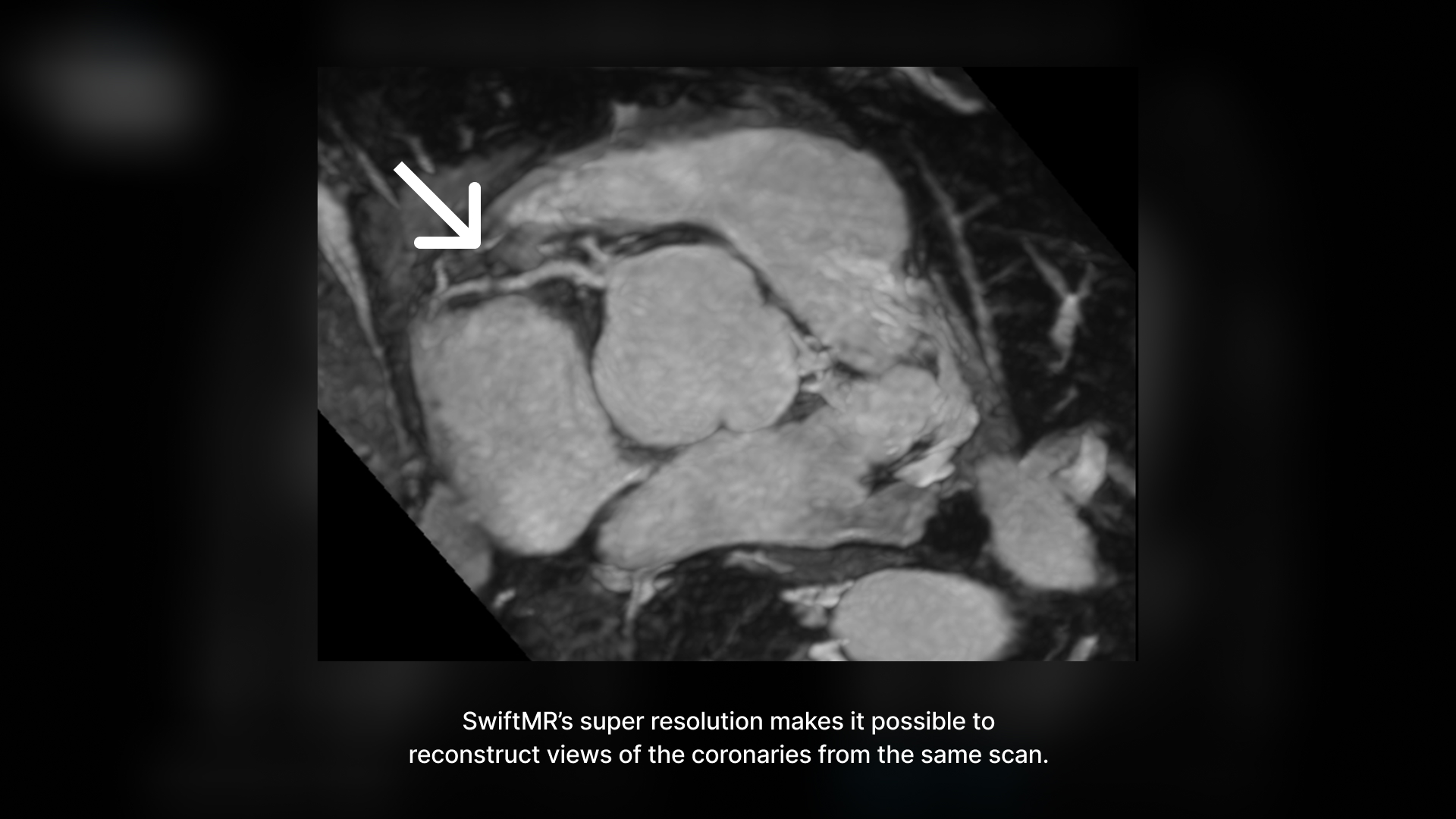
That is an important advantage of high-resolution 3D imaging: the value does not necessarily end with the primary aortic arch visualization. A volumetric dataset can provide additional anatomical perspectives through reconstruction and reformatting without requiring another dedicated acquisition.
Beyond image quality: Deep learning reconstruction unlocks faster CMR
Deep learning reconstruction is often discussed primarily in terms of image quality. In practice, its value can extend further.
When incorporated into an optimized acquisition strategy, SwiftMR can help imaging teams use acceleration more aggressively while producing the image quality required for clinical review.
In this case, the combination resulted in:
- 50% shorter starting acquisition time before triggering
- High-resolution, non-contrast visualization of the aortic arch
- 0.4 × 0.5 × 0.94 mm reconstructed resolution
- Image quality maintained in the accelerated dataset
- Additional coronary views reconstructed from the 3D acquisition
For complex, motion-sensitive examinations such as CMR, reducing the time required for individual acquisitions can have an impact beyond the sequence itself—helping make advanced imaging easier to incorporate into an already demanding examination.
Learn More
Explore more SwiftMR clinical images in the AIRS Medical image gallery.
For additional clinical examples or protocol discussions, contact Keaur Patel, BA, R.T.(R)(MR)(ARRT), Director of Customer Success.