Intracranial vessel wall imaging is technically demanding. The vessel wall is small, adjacent blood signal must be suppressed, and image quality depends on sufficient spatial resolution, SNR, and anatomic sharpness.
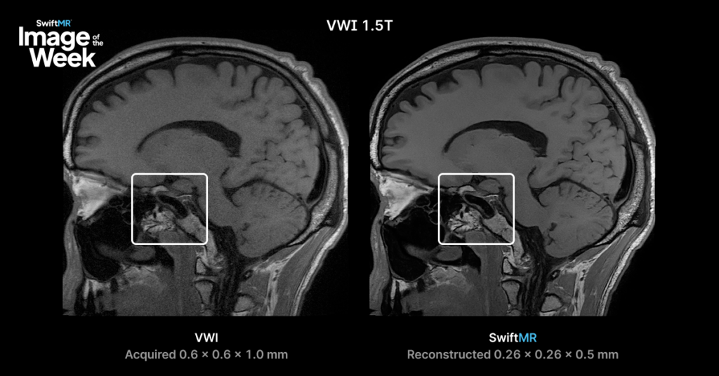
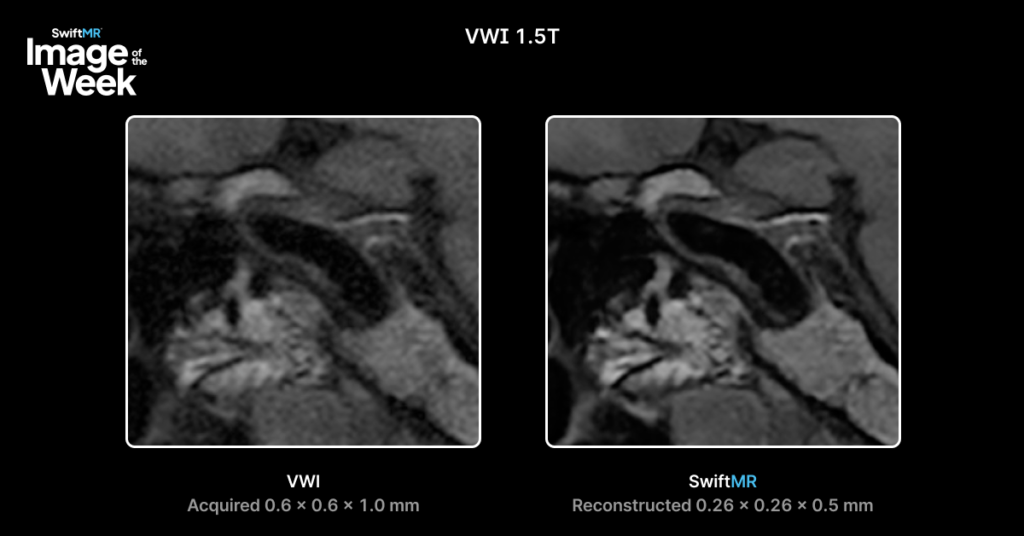
This clinical example compares a 1.5T intracranial vessel wall imaging acquisition with SwiftMR reconstruction.
The original image was acquired at 0.6 × 0.6 × 1.0 mm and reconstructed with SwiftMR to 0.26 × 0.26 × 0.5 mm, resulting in a smaller reconstructed voxel volume.
Areas of Interest
This comparison highlights differences in:
- Overall image sharpness
- Background noise
- Preservation of fine anatomic detail
- Multiplanar visualization
Because both images are from the same 1.5T vessel wall imaging acquisition, this comparison provides an opportunity to assess how SwiftMR reconstruction may influence image sharpness, reconstructed resolution, and review quality across planes.
Clinical Considerations
For radiologists, the value is the ability to evaluate sharper reconstructed detail and multiplanar views from the same 1.5T acquisition.
This is especially relevant for vessel wall imaging, where small structures and subtle findings often need to be reviewed with high spatial clarity.
Image Details
- Field Strength: 1.5T
- Application: Intracranial Vessel Wall Imaging
- Acquired Resolution: 0.6 × 0.6 × 1.0 mm
- SwiftMR Reconstructed Resolution: 0.26 × 0.26 × 0.5 mm
Comparison:
- Standard Acquisition
- SwiftMR Reconstruction
View the Dataset
To review the dataset, visit:
https://pacsbin.com/viewer/playlist/l_tmltNehjSb/review
Additional Resource
For additional clinical examples, visit the AIRS Medical clinical image gallery or contact Keaur Patel BA, R.T.(R)(MR)(ARRT), Director of Customer Success to discuss specific MRI protocols and imaging applications.