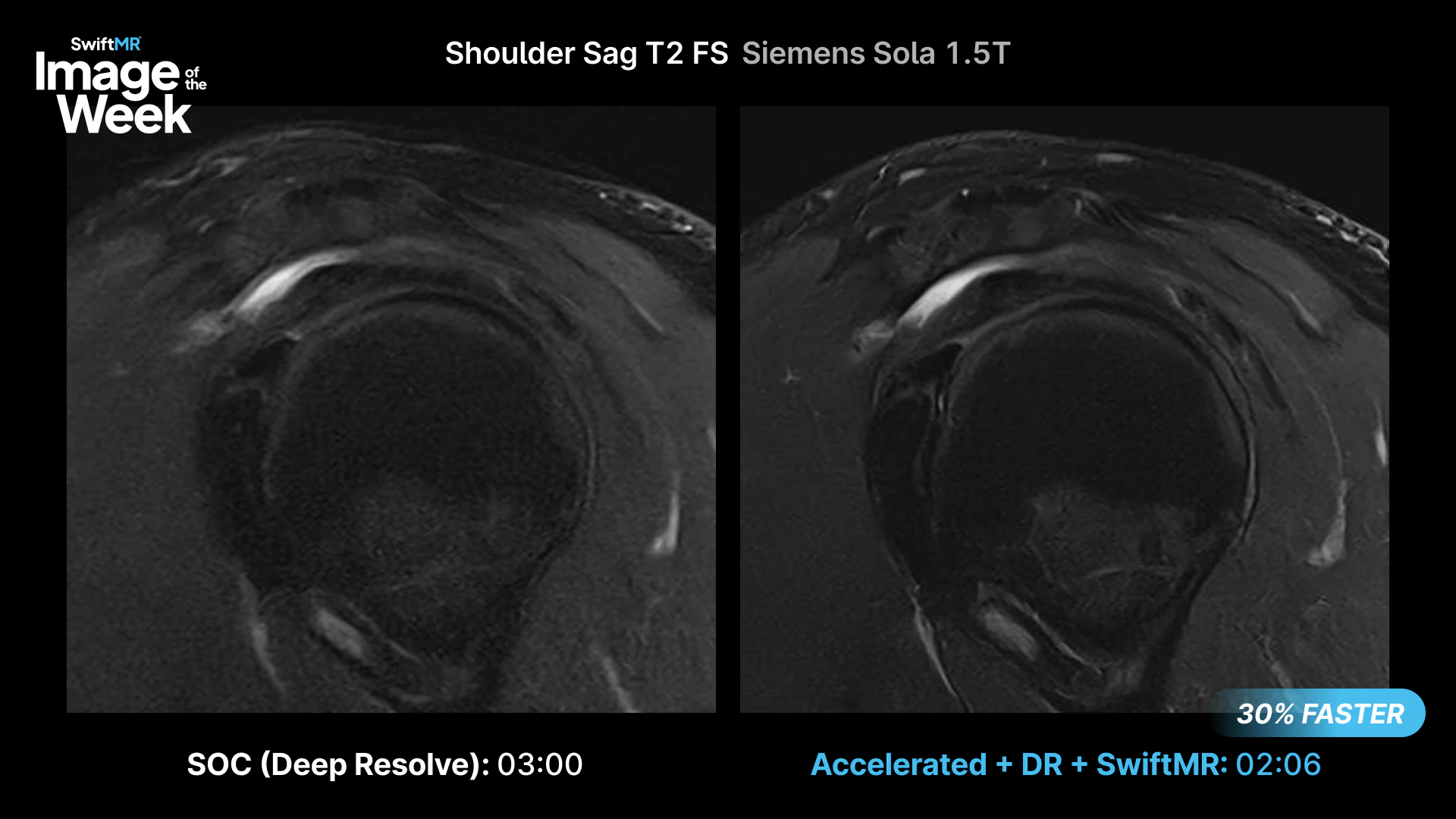
Sagittal T2 FS Shoulder: 3:00 to 2:06
This Sagittal T2 FS shoulder was acquired on a Siemens Sola 1.5T using Deep Resolve Boost.
| Acquisition | Scan time |
|---|---|
| Standard of care + Deep Resolve Boost | 3:00 |
| Deep Resolve Boost + SwiftMR | 2:06 |
With further acceleration and SwiftMR applied at the DICOM level after Deep Resolve Boost reconstruction, the sequence was acquired 54 seconds faster.
Fat-sat sequences in the shoulder can be particularly challenging because of off-isocenter positioning, the air-tissue interface at the deltoid, and the difficulty of achieving consistent shimming.
For this acquisition, combining Deep Resolve Boost with SwiftMR provides:
- Further noise reduction on an already-accelerated acquisition
- Clear muscle, tendon, and cortical margins
- Additional acceleration without replacing the OEM reconstruction
Deep Resolve Boost and SwiftMR operate at different stages of the imaging chain. Deep Resolve Boost supports acceleration during reconstruction in k-space, while SwiftMR applies image-domain denoising and super-resolution to the reconstructed DICOM images.
That combination can help support shoulder imaging where maintaining image quality may otherwise require additional scan time through techniques such as:
- Additional averages
- Tighter shim volumes
- Lower bandwidth
- Longer acquisitions to preserve SNR
Importantly, the additional image-domain processing was applied after the OEM reconstruction rather than replacing it.
In this acquisition, that resulted in a 54-second reduction on a single sequence, from 3:00 to 2:06.
Explore more clinical MRI examples
See additional SwiftMR clinical images across neuro, musculoskeletal, body and other MRI applications in the AIRS Medical Clinical Image Gallery.
Related reading: 7 Ways SwiftMR Improves OEM DL Reconstruction for Radiology Practices.
For additional clinical examples or protocol discussions, contact Keaur Patel, BA, R.T.(R)(MR)(ARRT), Director of Customer Success.