A 1:33 3D T1 for brain volumetry
Most volumetry protocols lean on an MPRAGE that runs 4 to 5 minutes. This week’s case is a 3D T1 SPACE acquired in 1:33 on a Siemens MAGNETOM Sola 1.5T, reconstructed with SwiftMR®, then segmented in SwiftSight®.
What to look for
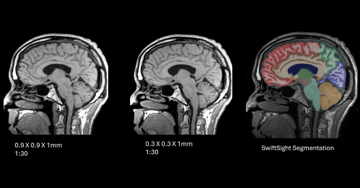
Left: the 1:33 acquisition as it came off the scanner (0.9 × 0.9 × 1 mm). Middle: the same data after SwiftMR reconstruction (0.3 × 0.3 × 1 mm). Right: SwiftSight’s segmentation of the SwiftMR images.
In the middle image, fine detail such as the cerebellar folia and the gray-white boundary along the cingulate is sharper. That’s the detail segmentation relies on, and it’s visible on a scan that took about a third of the usual time.
Time back on every brain exam
That’s roughly three minutes back per patient on a single series. Over a full day of brain exams, it’s an extra slot or a schedule that actually runs on time.
Explore more clinical MRI examples
See additional SwiftMR clinical images across neuro, musculoskeletal, body and other MRI applications in the AIRS Medical Clinical Image Gallery.
For additional clinical examples or protocol discussions, contact Anthony Rodenberg, BA, R.T.(R)(MR)(ARRT), Director of Clinical Programs.