Fetal cardiac cine is one of the most challenging CMR exams
With a heart roughly the size of a strawberry and a rapid rate of 120 to 160 bpm, fetal cardiac cine imaging presents severe technical constraints. Gating, fetal movement and maternal discomfort make it a difficult exam, ultimately leading to decreased image quality.
Under these circumstances, compromises are necessary: larger voxels preserve SNR, and shorter acquisitions can help mitigate motion artifacts. Even with an optimized protocol, standard acquisitions can result in grainy images with poorly defined myocardial borders.
In this case, SwiftMR® deep learning image reconstruction was combined with Northh Medical’s Smart-Sync Doppler ultrasound gating, which detects and synchronizes the fetal heartbeat with the MR system.
Image Details
| Scanner | Siemens MAGNETOM Aera 1.5T |
|---|---|
| Exam | Fetal cardiac cine |
| Gating | Northh Medical Smart-Sync Doppler ultrasound gating |
| Acquired resolution | 1.4 × 1.7 × 3.0 mm |
| SwiftMR reconstructed resolution | 0.26 × 0.26 × 3.0 mm |
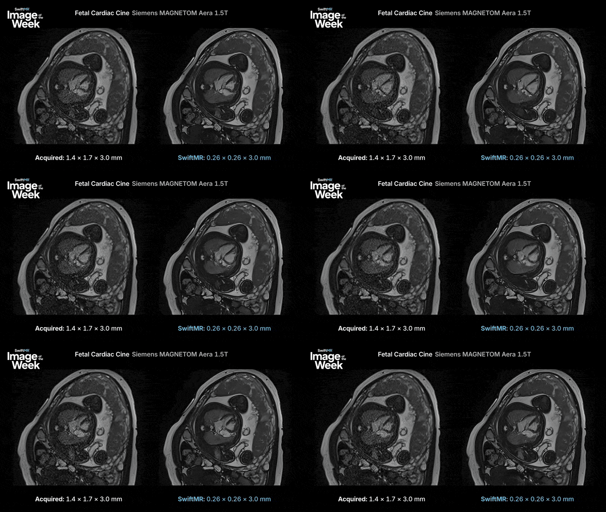
Left: conventional reconstruction. Right: the same acquisition processed with SwiftMR.
Look at the ventricular septum, valve leaflets, and the blood-myocardium boundary. Noise has decreased and the edges are sharper, which is exactly where fetal cardiac reads get uncertain.
Learn More
Explore more SwiftMR clinical images in the AIRS Medical clinical image gallery.
For additional clinical examples or protocol discussions, contact Anthony Rodenberg, BA, R.T.(R)(MR)(ARRT), Director, Clinical Programs.