When a patient has persistent leg pain and a lumbar spine MRI does not fully explain the symptoms, attention may turn beyond the spine itself to the lumbosacral plexus and peripheral nerves.
The lumbar plexus forms within the psoas muscle; the sacral plexus lies on the posterior pelvic wall over the piriformis. The major roots are easy to find. The smaller branches, like the obturator, pudendal and gluteal nerves, are not. The roots run obliquely before converging into the femoral, obturator and sciatic nerves.
Visualizing those pathways requires effective fat suppression, sufficient spatial resolution and enough signal-to-noise ratio to distinguish neural structures from surrounding tissue.
That combination places significant demands on the MR acquisition.
Why 3D MR neurography is challenging
Fat suppression is critical in lumbosacral plexus neurography because nerves must be distinguished from abundant surrounding fat within the pelvis.
Inversion-recovery fat suppression (STIR) is far less sensitive to B0 inhomogeneity than spectral methods, which matters over a field of view as large as the pelvis.
A 3D acquisition also allows anatomy to be evaluated beyond conventional axial images. The acquired volume can be reformatted along the oblique course of individual nerve roots, while maximum intensity projections (MIPs) can help visualize the plexus as neural structures extend toward larger peripheral nerves.
But there is a trade-off.
Techniques that rely on inversion recovery can reduce available signal, while small voxels cost SNR, and the long echo trains that keep scan time down add T2 blurring. The very characteristics that make 3D MR neurography valuable can also make the resulting images particularly sensitive to noise.
A 4-minute lumbosacral plexus MR neurography acquisition
Here’s a 3D SPACE lumbosacral plexus exam on a 3T Siemens MAGNETOM Vida.
| Scanner | Siemens MAGNETOM Vida 3T |
|---|---|
| Sequence | 3D SPACE lumbosacral plexus MR neurography |
| Scan time | 4:00 |
| Resolution | 1.1 × 1.4 × 1.4 mm |
| Display | 20 mm MIP |
One acquisition, processed two ways
What changes is the visualization.
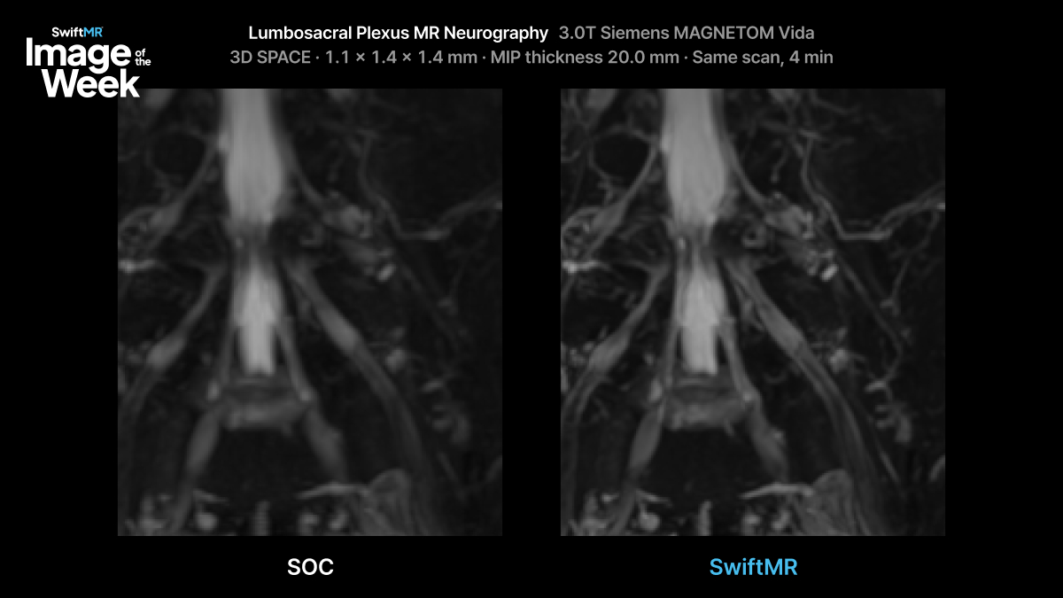
3D SPACE lumbosacral plexus MR neurography acquired on a 3T Siemens MAGNETOM Vida in 4:00 at 1.1 × 1.4 × 1.4 mm, shown with 20 mm MIP reconstruction. Conventional image shown alongside SwiftMR.
What changes with SwiftMR?
When the images are compared side by side, several differences become apparent.
The nerves also stand out more clearly against the background. A MIP keeps the brightest voxel along each path through the slab, so noise in the source images raises the background signal. With less noise going in, the background drops and the nerves separate from it.
Perineural tissue and branching architecture are more clearly visualized. Branches that fade into the background on the conventional MIP can be followed further on the SwiftMR image.
For an examination built around visualizing small, complex neural anatomy, those differences matter.
More clarity without adding acquisition time
Importantly, the improvement in visualization in this example does not require extending the scan.
The examination remains a 4-minute acquisition on the same 3T scanner.
That is particularly relevant for technically demanding sequences where simply increasing acquisition time to recover more signal may not be desirable. Longer acquisitions can increase the opportunity for patient motion while placing additional pressure on already constrained MRI schedules.
SwiftMR processes the images the scanner has already acquired, so the improvement comes without adding time to the exam.
For lumbosacral plexus MR neurography, where evaluation depends on following small neural structures through a complex anatomical background, clearer visualization can make the examination easier to interpret.
Explore more clinical MRI examples
See additional SwiftMR clinical images across neuro, musculoskeletal, body and other MRI applications in the AIRS Medical Clinical Image Gallery.
For additional clinical examples or protocol discussions, contact Keaur Patel, BA, R.T.(R)(MR)(ARRT), Director of Customer Success.