How Steinberg Diagnostic reached modern 3T image quality on its legacy MRI fleet with SwiftMR®
10 legacy Canon scanners running SwiftMR
50,000+ mRIs reconstructed with SwiftMR to date
4× increase in spatial resolution
Presented by Steinberg Diagnostic Medical Imaging at the RSNA 2025 annual meeting
President & Founding Partner, Neuroradiologist
Steinberg Diagnostic Medical Imaging
Trained at Brown, Stanford, and UCSF, Dr. Winkler was clinical director of the MRI Research Facility at UCSF before helping found SDMI. A neuroradiologist with nearly 100 peer-reviewed publications, he has spent years refining MRI sequencing across successive scanner generations and evaluating AI reconstruction solutions, and led SDMI's adoption of SwiftMR.
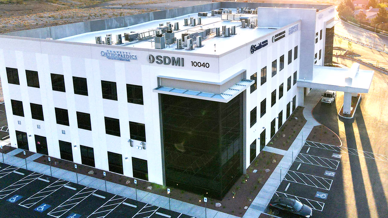
Steinberg Diagnostic Medical Imaging has served Las Vegas since 1988
Steinberg Diagnostic Medical Imaging (SDMI) operates 12 imaging centers across the Las Vegas Valley, providing MRI, CT, mammography, and a full range of diagnostic imaging services to Southern Nevada. Founded in 1988, it is one of the region's longest-established outpatient imaging providers.
The practice was built on a founding philosophy from Dr. Leon Steinberg: “If you practice great medicine, patients will follow.” Decades on, SDMI runs a high-volume MRI program on an aging fleet, and needed a way to keep image quality high without replacing the scanners behind it.
Why SDMI explored AI reconstruction
Two problems pushed SDMI to evaluate AI reconstruction. Both had to be solved without disrupting the radiologists and technologists who relied on the existing fleet every day.
The goal: improve image quality and extend the life of the legacy MRI fleet, getting more from the scanners SDMI already owned without a capital replacement cycle.
One solution had to meet five strict criteria
SDMI evaluated AI reconstruction against a clear set of requirements. The solution had to work everywhere, raise image quality, deploy easily, scale across the fleet, and prove itself against the alternatives. SwiftMR met every one, and won the head-to-head shoot-out.
All sequences, all body parts
Coverage across the full range of clinical exams, not a narrow subset.
Improve image quality
A genuine step up in image quality, not just faster acquisition.
Easy on-site implementation
Deployable on the existing fleet without disruptive installation.
Easy to scale
Roll out across many scanners and sites without added complexity.
Win a competitive shoot-out
Outperform other reconstruction solutions in a direct, head-to-head evaluation.
SwiftMR delivered on image quality
The deciding factor was image quality. Across SDMI's exams, SwiftMR combined three improvements that, together, brought legacy scanners up to a modern 3T look.
Noise reduction
Cleaner images with a markedly higher signal-to-noise ratio, even on older 1.5T systems.
4× spatial resolution
Sharper anatomical detail and finer structures resolved across sequences and body parts.
Artifact reduction
Fewer truncation, susceptibility, and motion artifacts, for more natural-looking studies.
Image gallery
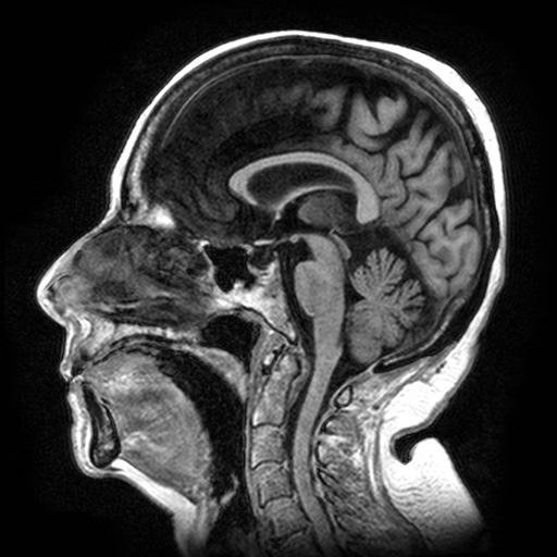
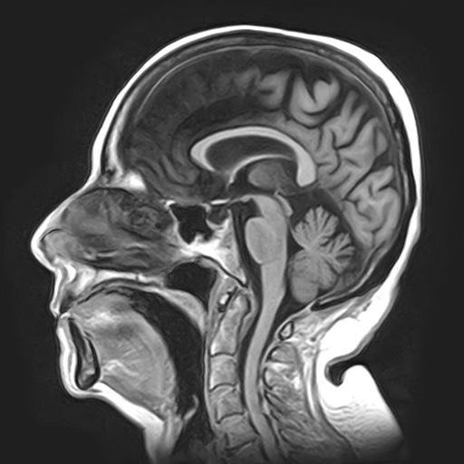
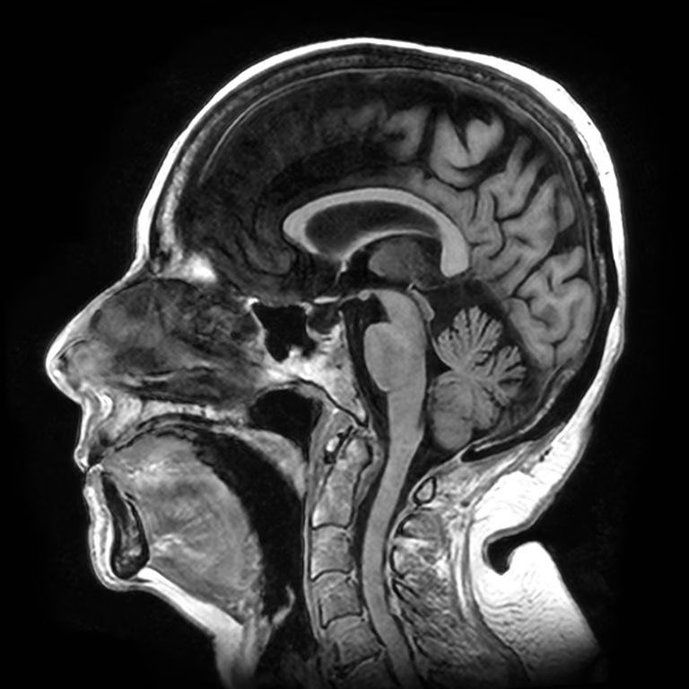
Comparisons on SDMI's Canon scanner fleet. Baseline and conventional images are shown at their acquired resolution. Individual outcomes vary by scanner model, field strength, pulse sequence, and clinical protocol.
SwiftMR brought modern image quality to the entire fleet
Deployed across 10 Canon scanners
SwiftMR rolled out fleet-wide, with no need to replace any legacy systems.
No workflow changes
Technologists kept their existing routines. SwiftMR slots into the established imaging workflow.
Image quality gains everywhere
Consistent gains across every sequence and body part, not just a handful of protocols.
Legacy systems, extended
Modern 3T-class image quality from scanners SDMI already owned, lengthening their useful life.
“SwiftMR gives our legacy scanners modern 3T image quality: higher signal-to-noise, higher resolution, and fewer artifacts, with no workflow changes and no need to replace our scanners.”
Mark L. Winkler, MDPresident & Founding Partner, Steinberg Diagnostic Medical Imaging
Bring modern image quality to your legacy scanners
SwiftMR's deep learning technology reconstructs MRI scans, delivering sharper, cleaner images on the equipment you already own. Providers like SDMI have used it to raise image quality across an aging fleet and extend the life of legacy systems, with no new hardware and no workflow changes. Talk to AIRS Medical about what SwiftMR can do for your scanners.
SwiftMR® is FDA 510(k) cleared as an AI-powered MRI image enhancement solution intended for noise reduction and increased image sharpness on non-contrast enhanced MRI images in DICOM format. Supported body parts, pulse sequences, field strengths, patient populations, and MRI manufacturers may vary by country.
Content adapted from a presentation by Mark L. Winkler, MD, at the RSNA 2025 annual meeting, “Enhancing Image Quality Across Legacy MRI Scanners.” Image comparisons show baseline acquisition and conventional reconstruction versus SwiftMR on SDMI’s Canon scanner fleet. Individual outcomes may vary by scanner model, field strength, pulse sequence, and clinical protocol; individual practices should not expect identical results.
Quotes and data published with Steinberg Diagnostic Medical Imaging’s permission. This page is intended for healthcare professionals.
© 2026 AIRS Medical Inc. SwiftMR® is a registered trademark of AIRS Medical Inc.