Eight Denoising Levels: What Changes—and What Doesn’t?
There is no single definition of a good MRI image.
Resolution, contrast, noise, and texture all contribute to image quality, but their relative importance changes depending on the anatomy, sequence, and clinical question.
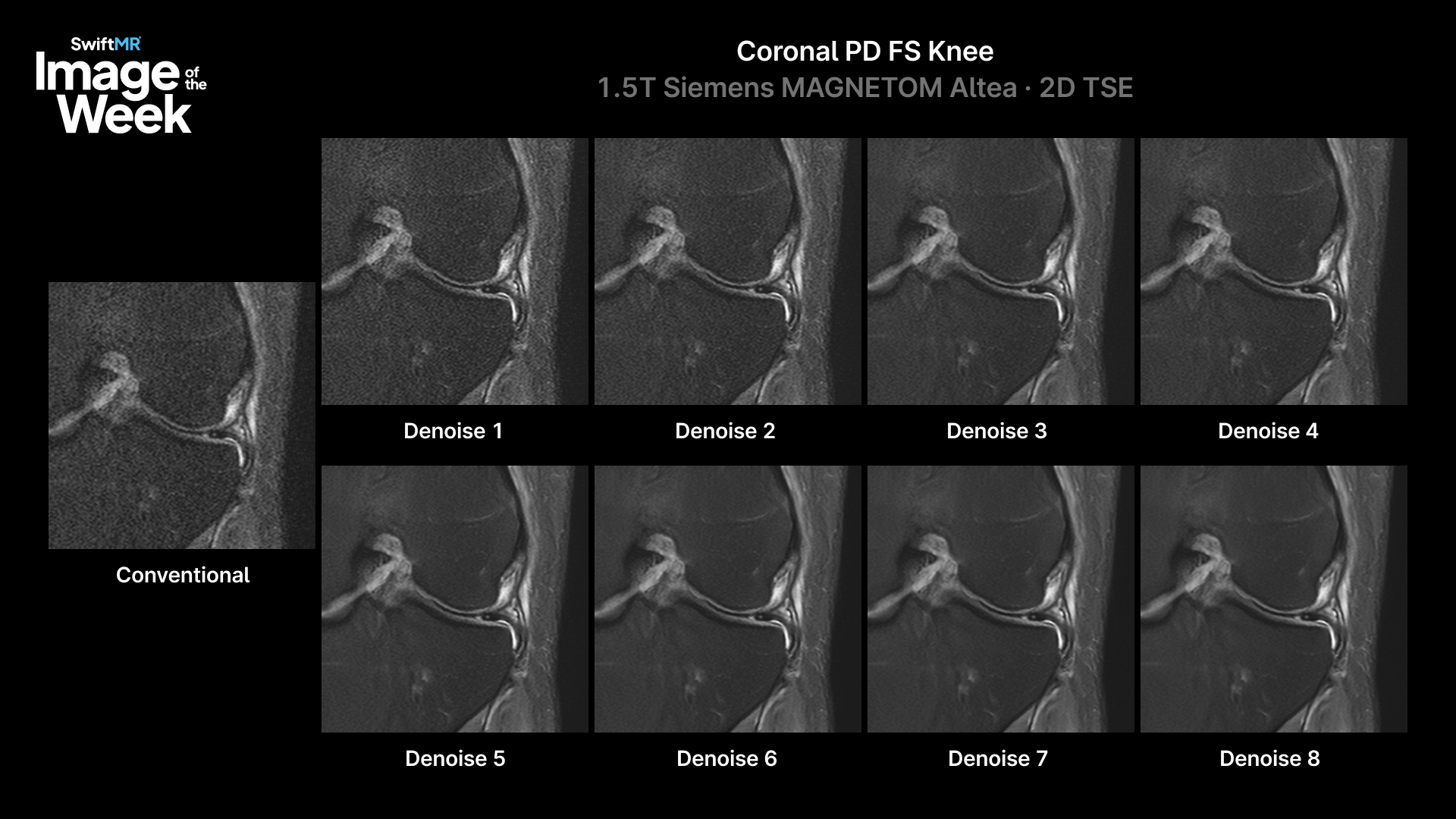
This knee MRI provides a useful example of what happens when the same sequence is reconstructed across eight different denoising levels.
Clinical example
| Scanner | Siemens MAGNETOM Altea 1.5T |
|---|---|
| Sequence | Coronal PD FS (TR 3510 / TE 41) |
| Scan time | 52 seconds |
| Acquired resolution | 0.45 × 0.57 × 3.0 mm |
| Reconstruction resolution | 0.16 × 0.16 × 3.0 mm |
| SwiftMR denoising levels evaluated | 1–8 |
| Radiologist selection | Level 6 |
What are we actually changing when we denoise an MRI?
Denoising reduces image noise, but the objective isn’t simply to produce the smoothest possible image.
Radiologists still need the image characteristics required to evaluate the anatomy in question—including tissue contrast, edges, fine morphology, and natural image texture.
That balance changes with the clinical task.
A small meniscal tear, for example, can depend on visualization of subtle morphology. Early ischemic change is much more dependent on contrast. Increasing spatial resolution alone doesn’t necessarily add the same value to both examinations.
The same principle applies to reconstruction.
One reconstruction setting doesn’t fit every sequence
MRI practices routinely use protocols across neuro, MSK, body, and vascular imaging. Within those protocols, individual sequences are designed to answer different clinical questions.
A reconstruction optimized for one sequence may therefore be less appropriate for another.
This is why SwiftMR provides eight denoising levels and adjustable reconstruction resolution that can be configured at the sequence level.
The objective is not to identify one universally “best” reconstruction setting. It is to give imaging teams the ability to match reconstruction characteristics to the sequence and clinical task.
What happens across the eight denoising levels?
Watch the progression from Level 1 through Level 8 in this knee MRI.
At the lower levels, the reduction in noise is readily visible.
Then look closely at Levels 5 through 8.
The differences become much more subtle. In this sequence, the effect of increasing denoising begins to plateau rather than continuing toward an increasingly smooth, artificial appearance.
The radiologist reviewing this case selected Level 6.
That doesn’t mean Level 6 is the correct setting for every knee sequence—or even every radiologist.
That’s the point.
More denoising isn’t necessarily better
The goal with denoising is to reduce noise while preserving the image characteristics needed for interpretation. Once the effect begins to plateau, additional denoising may provide little additional value.
The optimal balance can vary by anatomy, sequence, and clinical question—which is why reconstruction should be tunable at the sequence level rather than fixed across an entire MRI protocol.
The radiologist chose Level 6. Which one would you read?
Learn More
To learn more, visit the SwiftMR product page.
Explore more SwiftMR clinical images in the AIRS Medical image gallery.
For additional clinical examples or protocol discussions, contact Keaur Patel, BA, R.T.(R)(MR)(ARRT), Director of Customer Success.