
Finishing a head MRA acquisition doesn’t always mean it’s ready for interpretation.
The MR technologist may still need to open the dataset, define the reconstruction volume, isolate the vessels, select projection angles, create spin and tumble series, label the images, and send the completed reconstructions to PACS.
AIRS Medical’s latest SwiftMR update automates those steps with FDA-cleared Auto Cut MIP. It performs vessel segmentation and generates the standard and vessel-focused MIP views used for time-of-flight MRA Head. Rather than building each series manually, technologists receive a complete, consistently labeled set of reconstructions that routes directly to PACS.
The result is more than five minutes of technologist time saved per eligible examination, with more consistent output across technologists, scanners, and imaging locations.
For imaging organizations, the benefits extend beyond automated image creation:
- Standardized, consistently labeled vessel views for radiologists
- Less technologist-to-technologist variability
- Fewer back-and-forth requests between radiologists and technologists
- Faster study delivery to PACS with fewer post-processing delays affecting the daily schedule
- A scalable, vendor-neutral workflow across MRI fleets
What’s a MIP?
MIP stands for maximum intensity projection.
It’s a method for collapsing a 3D imaging dataset into a 2D view by displaying the highest-intensity signal along each line of sight.
In time-of-flight MRA, flowing blood appears bright. MIP processing uses that contrast to make the vessels stand out from the surrounding anatomy.
One projection often isn’t enough, because vessels at different depths overlap when flattened onto a single plane. That’s why MRA Head workflows use rotating spin and tumble series, plus focused views of specific vascular territories, so an overlap hidden at one angle separates out at another.
What SwiftMR Generates
SwiftMR automatically segments the vessels and creates the expected head MRA reconstructions.
For MRI Technologists: 5+ Minutes Saved per Head MRA
SwiftMR eliminates manual MIP post-processing from the technologist workflow, including vessel cuts, projections, series labeling, and routing.
The process is fully automated, with completed images sent directly to PACS without additional MRI technologist interaction. This can save more than five minutes per eligible MRA TOF head examination, improve study-to-read time, and potentially support faster report turnaround.
When used alongside SwiftMR scan acceleration, technologists also benefit from reduced acquisition time—giving them more time to stay on schedule and focus on patient care.
For Radiologists: Standardized MRA Output Across Scanners
SwiftMR delivers consistent vessel segmentation, MIP post-processing, and series labeling regardless of technologist skill level.
Radiologists receive reliably labeled combined bilateral carotid, separate left and right carotid, and vertebrobasilar views, with higher SNR and vessel conspicuity across MRI scanner makes and models. This reduces tech-to-tech variability and limits back-and-forth requests for missing or differently processed reconstructions.
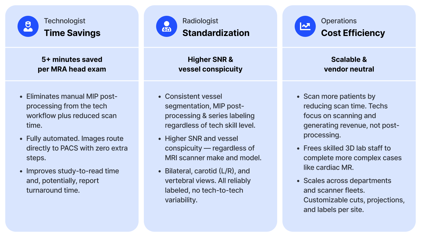
For Imaging Operations: Scalable, Vendor-Neutral Efficiency
SwiftMR reduces routine post-processing time so technologists can stay focused on scanning and maintaining the daily schedule rather than manually generating MIP views.
The automated workflow also frees skilled 3D lab staff to focus on more complex cases, such as cardiac MR.
Because SwiftMR is vendor-neutral, standardized cuts, projections, and labels can be customized by site and scaled across departments, scanner fleets, and imaging locations.
MIPs still support, not replace, source-image review
Automated MIP generation does not replace the source dataset or the radiologist’s clinical judgment.
The source images remain available for review and may provide information that is less apparent on a projection image, particularly where vessels overlap or flow-related effects are present.
SwiftMR handles the repetitive reconstruction work. The radiologist still interprets the complete examination.
See SwiftMR Auto Cut MIPs in Action
FDA-cleared automation for standardized head MRA post-processing.
FDA 510(k) database: SwiftMR K261349, dated July 24, 2026.