Twelve minutes of holding still, for a patient who can’t
Ask someone with essential tremor to stay perfectly still for twelve minutes and twenty-four seconds. That’s not a thought experiment. It’s the length of one scan in the workup before deep brain stimulation surgery, and it’s the scan that matters most.
Deep brain stimulation treats tremor, Parkinson’s disease and dystonia by placing a thin electrode into a specific structure deep inside the brain and delivering steady electrical pulses through it. The therapy can be remarkable. It also depends entirely on the electrode ending up in the right place, and the targets are small, buried in the middle of the head, and surrounded by structures you very much do not want to hit.
Which makes the imaging beforehand less like taking a picture and more like drawing a map someone is going to navigate by.
The problem with the middle of the brain
Most brain MRI is good at telling you where the gray matter is and where the white matter is. Gray matter is where the processing happens. White matter is the wiring that connects it.
The trouble is that the deep structures a surgeon targets sit in a crowded neighborhood, and on a normal scan they blend into their neighbors. You can see roughly where things are. You cannot see the borders between them, and the borders are the whole point.
There’s a sequence built for exactly this. FGATIR is set up so the white matter goes dark instead of bright. Turn down the wiring and the deep gray structures stand out from it, along with the thin sheets of wiring that thread between them. Those thin sheets are the landmarks. The surgeon uses them the way you’d use a street to tell one block from the next.
What the surgeon is actually looking for
The target depends on the patient and the condition, and each one comes with its own hazards nearby.
For the globus pallidus, a common target in Parkinson’s and dystonia, the surgeon needs to see a thin sheet of white matter that separates the target from the structure right beside it. Just as important is what surrounds it. The optic tract, which carries vision, runs underneath. Stimulate too close to it and the patient sees flashes of light. The internal capsule, which carries movement and speech signals, runs alongside. Stimulate too close to that and you get muscle pulling and slurred speech, which are the side effects that most often limit how much therapy a patient can tolerate. This sequence shows the target and the hazards in the same image.
For the VIM, the usual target for tremor, the difficulty is that the structure itself is nearly invisible on ordinary scans. It has no real edge to find. Surgeons navigate to it by measuring from nearby landmarks, and white-matter-nulled sequences like this one are what make those landmarks visible. Newer work suggests the target itself can sometimes be seen directly.
For the subthalamic nucleus, another common Parkinson’s target, this sequence is a supporting player rather than the star. Distinguishing that structure from its neighbor is a different imaging problem, better solved by other sequences. Centers doing this work use several sequences together, not one.
Why the length of the scan is the problem
Fine detail is the first thing lost when a patient moves. The overall picture usually survives. The thin lines and subtle edges, the exact things this scan exists to show, do not.
And moving is what this group of patients does. Tremor, rigidity, dystonia. In many protocols the patient is deliberately scanned off their medication, meaning at their symptomatic worst, because that’s the state the surgery is meant to address. Then we ask them to lie motionless for over twelve minutes for the single most detail-dependent sequence in the exam.
If it fails, the answer is to do it again. Same patient, less patience, more movement the second time.
Why the strength of the magnet matters
MRI scanners come in different field strengths. 3T is roughly twice as strong as 1.5T and generally gives more signal and finer detail, which is why planning scans have traditionally been done there.
After surgery, though, the patient has metal hardware in their head, and that changes the rules. Implanted devices carry safety labeling that spells out the conditions under which they can be scanned. Some current devices are approved for both 1.5T and 3T. Others are approved at 1.5T only. Either way the labeling limits how much radiofrequency energy the scan can deposit, which constrains what the protocol can do even when the scanner itself is allowed.
Add to that the hospitals whose own policies are stricter than the manufacturer’s, the patients who already have another implant restricting them, and the sites that simply don’t own a 3T. For a large share of these patients, 1.5T isn’t the fallback option. It’s the only option, which means the image quality has to be there.
What changed here
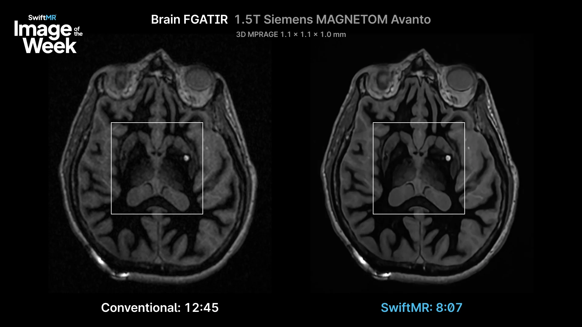
This case was a brain FGATIR on a Siemens MAGNETOM Avanto 1.5T, at the same resolution either way.
| Scanner | Siemens MAGNETOM Avanto 1.5T |
|---|---|
| Sequence | Brain FGATIR |
| Conventional acquisition time | 12:24 |
| SwiftMR acquisition time | 8:07 |
| Time reduction | 35% faster |
| Resolution | 1.1 × 1.1 × 1.0 mm (both) |
The conventional scan took 12:24. With SwiftMR, 8:07. Four minutes and seventeen seconds faster, about 35% less time, on the scan where lying still is the hardest part.
Four minutes isn’t a scheduling statistic on this exam. It’s the difference between a study the surgical team can plan from and a study that has to be repeated.
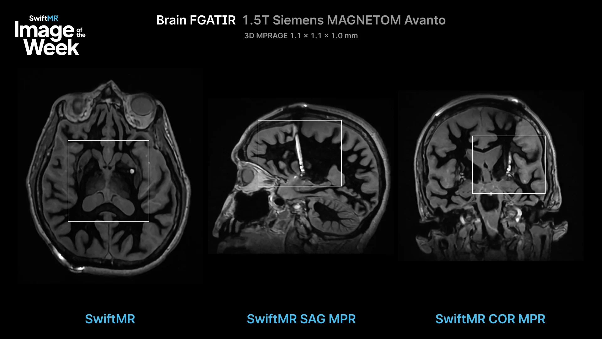
There’s a second benefit that’s easy to miss. Surgical planning and post-op assessment doesn’t happen only in the plane the scan was taken in. The team looks at the same data sliced along the path the electrode will travel, and those reconstructed views are only as good as the data behind them, including in the direction between slices. SwiftMR improves resolution in that direction too, not just within each slice, and builds those reconstructed views automatically from the improved images. The views the surgeon actually plans in get the same benefit as the one that was scanned.
Learn More
Explore more SwiftMR clinical images in the AIRS Medical clinical image gallery.
For additional clinical examples or protocol discussions, contact Keaur Patel, BA, R.T.(R)(MR)(ARRT), Director of Customer Success.