Wrist MRI leaves little room for compromised image quality. The anatomy is compact, the intrinsic ligaments are thin, and clinically important abnormalities can be subtle. Scaphoid fractures, in particular, often stay occult on initial radiographs, which is why MRI or CT is recommended when clinical suspicion persists despite negative radiographic findings.
For imaging centers using a low-field open system such as the Fujifilm APERTO Lucent 0.4T, the question isn’t whether the scanner can image the wrist. It’s whether it can deliver enough signal and spatial resolution to resolve that anatomy.
That question becomes particularly apparent with STIR added to the mix.
STIR provides robust fat suppression and makes fluid-sensitive findings, including marrow edema associated with occult fracture, more conspicuous. But inversion recovery also comes with a signal penalty. On 0.4T systems, where intrinsic SNR is already lower, fat nulling can make noise more visible and softens structural margins.
So you’re left with the familiar trade-off: keep the open low-field scanner and accept reduced image quality, or replace or upgrade the hardware at considerable cost.
In this case, there was a third option.
Improving Low-Field Wrist MRI with SwiftMR
The comparison shown here was acquired on a Fujifilm APERTO Lucent 0.4T open MRI system.
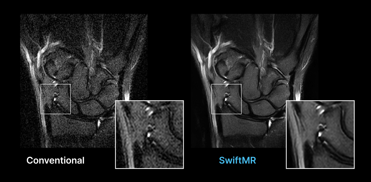
SwiftMR applies deep-learning denoising and super-resolution to the acquired STIR image data.
In the displayed comparison, visible noise is reduced and the margins of the carpal bones and surrounding soft-tissue structures appear more clearly defined.
The fat-suppressed contrast of the original STIR acquisition remains preserved. Nothing is reacquired, and the image isn’t converted into another sequence. SwiftMR recovered detail from what the acquisition already captured.
Clinical Relevance of AI-Reconstruction
Cleaner STIR images give radiologists a clearer basis for assessing marrow and soft-tissue abnormalities. Field strength still matters, but it’s no longer the only factor setting how much a low-field study can show.
Learn More
Explore the AIRS Medical clinical image gallery.
For protocol details or additional wrist MRI examples, contact Keaur Patel, BA, R.T.(R)(MR)(ARRT), Director of Customer Success.