Less Noise.Read with Confidence.
MRI doesn’t always deliver the image quality you want. Noise, challenging protocols, motion, and acquisition constraints can make images hard to interpret with confidence. SwiftMR® uses AI reconstruction to help improve image quality and preserve clinically relevant detail.
Clinically evaluated • Peer-reviewed evidence • Works within your PACS workflow
See More. Read Clearly.
See examples of how SwiftMR reduces noise and visual distraction across anatomy, scanners, and clinical applications.
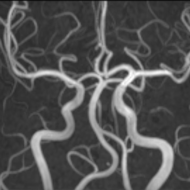
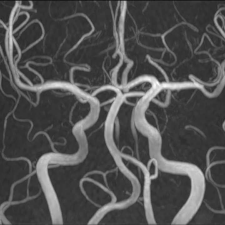
BRAIN MRA · 3.0T Siemens MAGNETOM Skyra
Reduced noise · Same acquisition
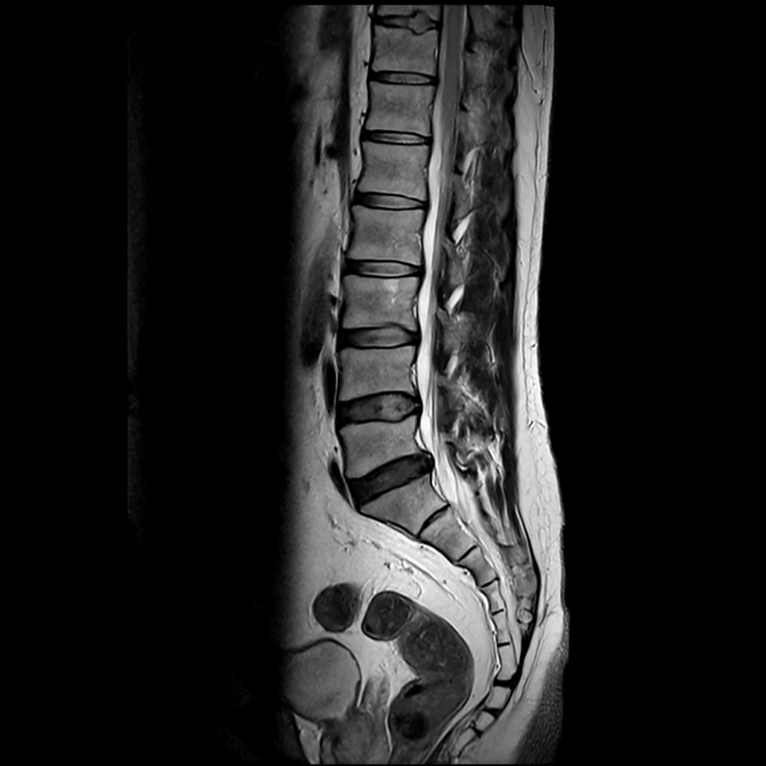
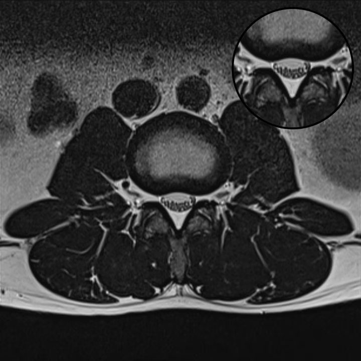
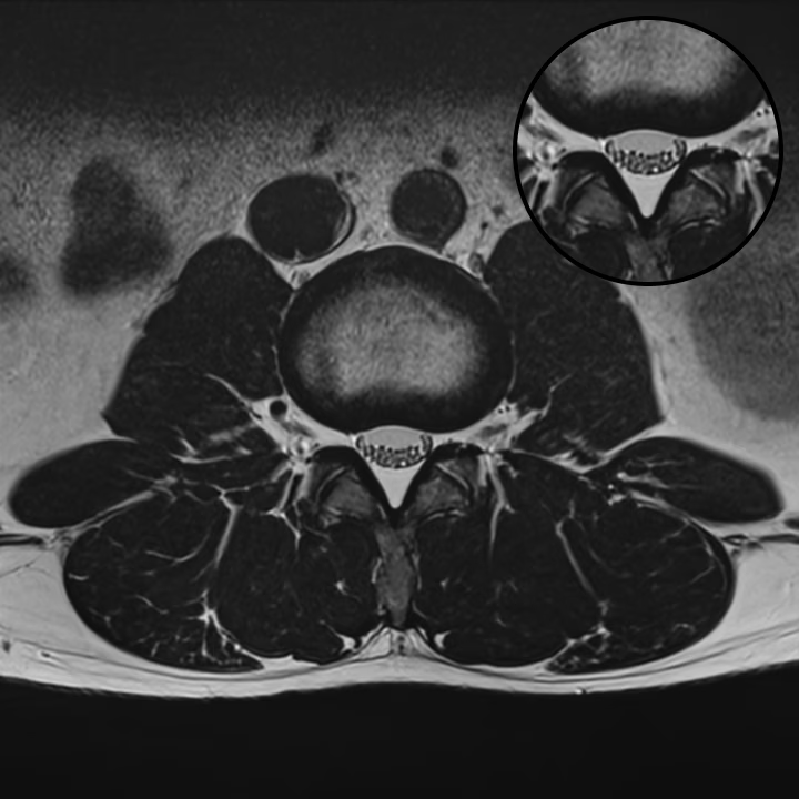
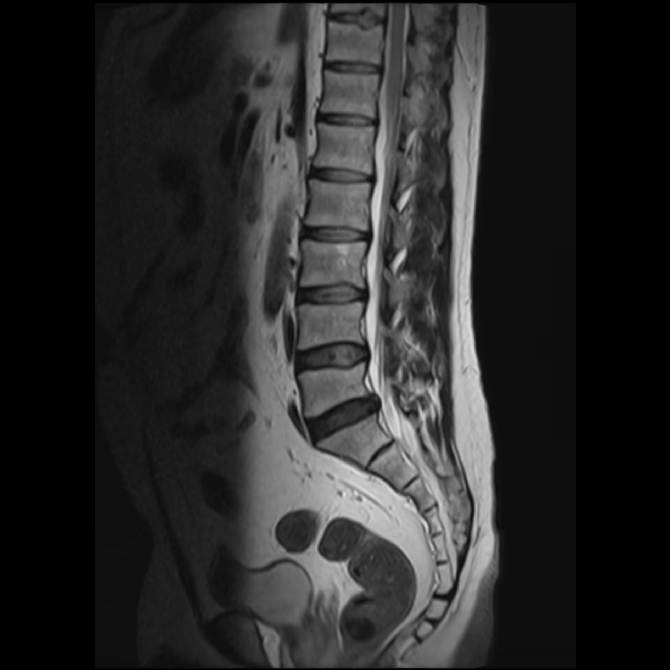
L-SPINE T2WI · 1.5T Siemens MAGNETOM Essenza
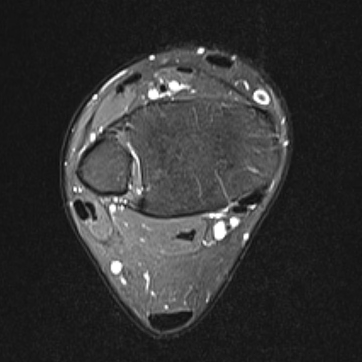
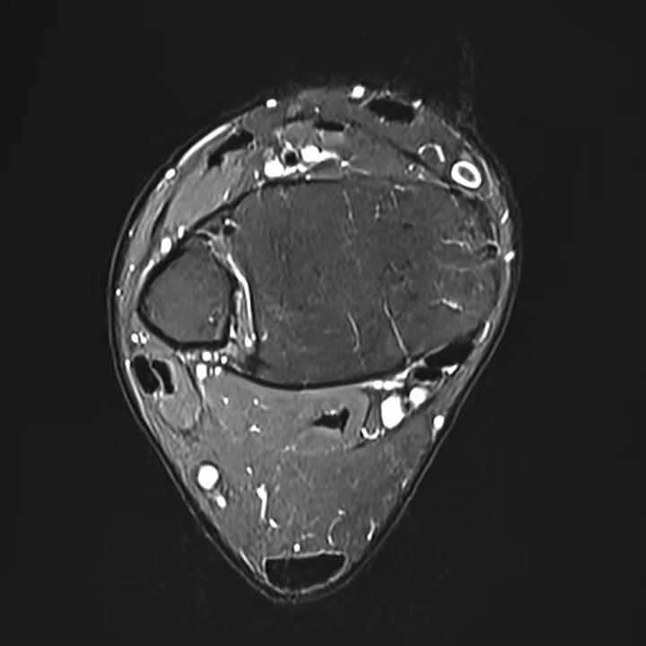
ANKLE PDWI FS · 1.5T Siemens MAGNETOM Essenza
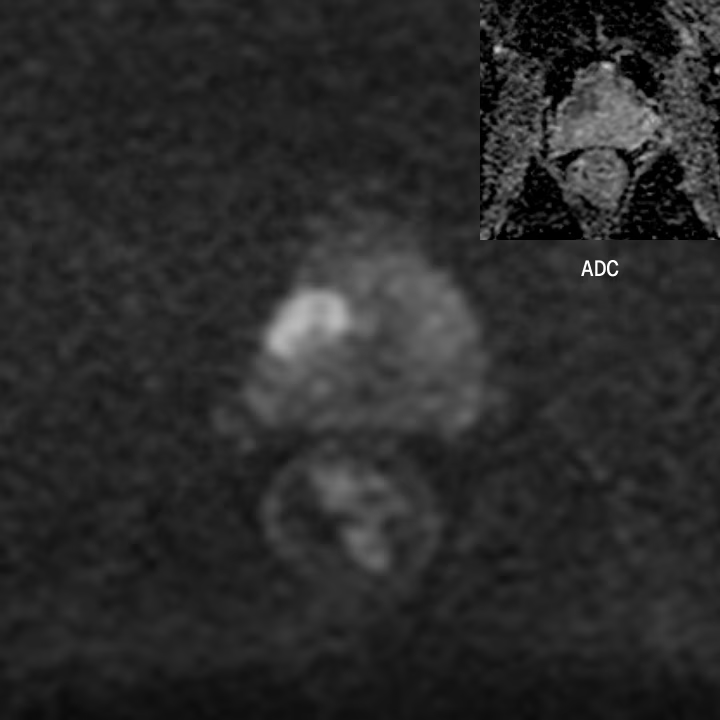
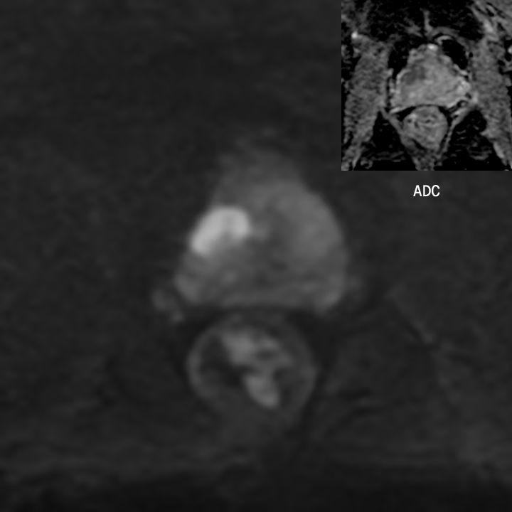
PROSTATE DWI · 3.0T GE DISCOVERY MR750w
2-CHAMBER CINE · 11 second breath hold
2-Chamber cine · 4 second breath hold
More Detail. Less Distraction.
SwiftMR reduces noise while preserving natural image texture, tissue contrast, and anatomical detail, across scanners, sequences, and protocols.
Improves the visibility of fine anatomical structures and supports higher-resolution imaging.
Preserves contrast between adjacent tissues while reducing noise.
Reduces image degradation and visual distraction without requiring reacquisition.
Supports clearer visualization of subtle findings against surrounding tissue.
No New Viewer. Same Workflow.
Images are processed in the background and returned to PACS, so they can be reviewed within your existing reading workflow.
Accelerated sequences run on your existing scanner. To the MRI, SwiftMR looks like a PACS server: standard DICOM transfer, with no software installed on the equipment.
Images are de-identified at the gateway (HIPAA Safe Harbor) and processed in the cloud, typically in under 2 minutes. Cloud data is deleted within 24 hours.
Enhanced images are re-matched to the patient and stored to PACS automatically. By the time the patient is off the table, results are on the worklist.
You read in your existing viewer: no separate application, no manual upload, just clearer images in the same workflow.
MIP Series Generated Automatically
SwiftMR automatically creates standardized MIP views from the source acquisition and returns them to PACS, reducing manual post-processing before interpretation.
Proven Performance AcrossKey Clinical Areas
SwiftMR has been evaluated across brain, spine, musculoskeletal, and neurovascular imaging, with peer-reviewed studies published in leading radiology journals.
39.4% shorter scan time with improved image quality
In a prospective study of 150 participants scanned on three 3T MRI systems from different vendors, SwiftMR was applied simultaneously to routine 2D spin-echo and 3D gradient-echo sequences. Accelerated MRI demonstrated improved overall image quality and structural delineation compared with conventional MRI.
Korean Journal of Radiology · 2025
Choi KS, Park C, Lee JY, et al.
32.3% shorter acquisition time with maintained diagnostic performance
In a prospective study of 50 patients undergoing lumbar MRI at 1.5T, accelerated images reconstructed with SwiftMR demonstrated similar or better image quality than standard MRI. Radiologist sensitivity and specificity for major degenerative lumbar spine findings were not significantly different.
41% shorter scan time with comparable diagnostic performance
In a prospective study of 45 patients across three 3T MRI systems from Philips, Siemens, and GE, accelerated knee MRI reconstructed with SwiftMR produced higher SNR and CNR while maintaining comparable overall image quality and lesion detection. Images were independently evaluated by three musculoskeletal radiologists.
Faster, more confident large-vessel occlusion assessment
In a 520-patient study, SwiftMR-enhanced 4D-TWIST MRA improved image quality. In patients evaluated for suspected acute ischemic stroke, it also increased radiologist confidence and reduced the time required to assess large-vessel occlusion compared with the original 4D-TWIST images.
Kim BK, You S-H, Kim B, Shin JH
Published in leading peer-reviewed radiology journals
Evaluate SwiftMR on Your Clinical Cases
You don’t have to take our word for the image quality. During the one-month free trial, SwiftMR is validated on your own protocols and pathology. Your radiologists make the call at every step before anything changes in routine use.
Select representative protocols and cases
Choose exams that reflect your clinical routines and questions.
Run conventional, accelerated, and SwiftMR sequences.
Review with the relevant radiologists
Assess diagnostic confidence and overall image quality.
Refine and approve incrementally
Collaborate and adjust parameters to scale with confidence.
Frequently Asked Questions
No. SwiftMR is trained on pairs of images showing identical anatomy at different noise levels, so the algorithm removes noise patterns while preserving anatomical structures, including lesions. Every published peer-reviewed SwiftMR study to date has demonstrated at least non-inferiority to standard imaging in image quality and diagnostic performance.
You can also verify this directly: during an evaluation, radiologists review representative pathology side by side, assessing anatomy, lesion visibility, contrast, image texture, and artifacts before approving any sequence for routine use.
SwiftMR is designed to restore natural, consistent image texture, and peer-reviewed reader studies report no degradation in image quality. Sharpness and denoising levels are tuned by sequence based on your radiologists' feedback until images read the way your team expects.
During an evaluation, image texture, edge appearance, and anatomical detail are reviewed across representative sequences and anatomies before anything enters routine use.
Yes. AIRS Medical provides a one-month free trial, and accelerated sequences can initially be added alongside the existing standard-of-care examination, so nothing changes until your team is satisfied. Accepted sequences are then introduced incrementally.
No. Protocol selection remains with the radiologist and MRI team. SwiftMR focuses on reconstruction and enhancement of acquired MRI images, and runs as a stand-alone system that cannot install software on or affect the scanner in any way.
SwiftMR is compatible with all MRI vendors and scanner models, regardless of their age or magnet type, at field strengths from 0.25T to 3.0T. This includes open and upright MRI systems.
If you're looking to implement SwiftMR in MRI vendors other than GE, Siemens, Philips, Canon (Toshiba), Fujifilm (Hitachi), and Fonar, please reach out to our team!
Yes. SwiftMR is FDA-cleared to work in conjunction with vendor deep learning reconstruction solutions such as GE AIR Recon DL, providing further acceleration on top of your current parameter settings. It can also extend reconstruction coverage to sequences, anatomies, or scanners the OEM solution does not cover.
Bring Greater Clarity to the MRI Read
Join radiology teams already saving time and reading with greater confidence.
Plan a Clinical Evaluation
Talk to our clinical team about your center's cases and challenges. Bring your own images to try SwiftMR.
Start Your Free Trial
No commitment. Experience SwiftMR for 30 days in your own environment.