How Mag-Medica increased daily MRI capacity by 20% without replacing its scanner
On its Siemens Avanto 1.5T, SwiftMR reduced average acquisition times by 39.5% while producing sharper, less noisy images across neuro, musculoskeletal and body imaging.
Radiologist, Mag-Medica · Novi Sad, Serbia
One aging scanner, a growing caseload
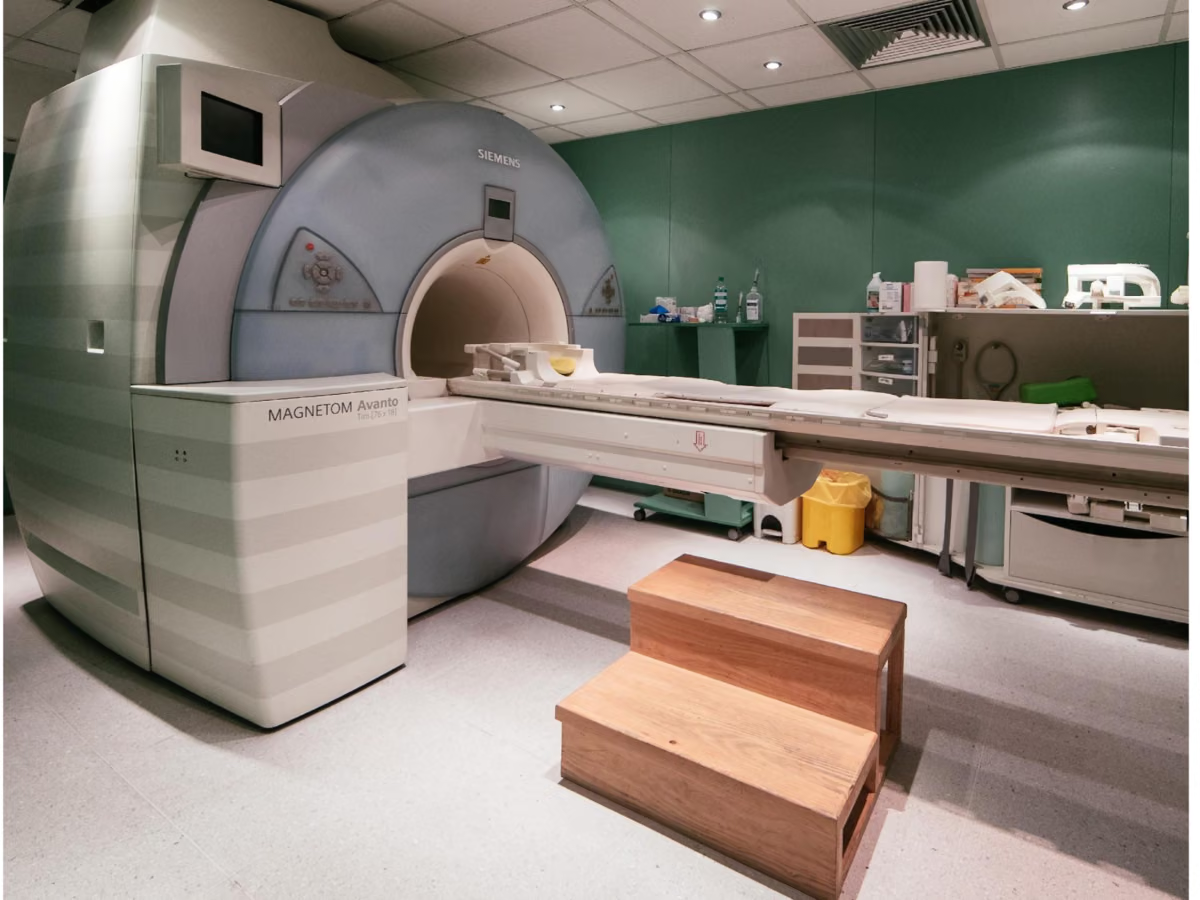
Mag-Medica is a single-center imaging clinic in Novi Sad, Serbia. Its entire MRI service runs through one 21-year-old Siemens Magnetom Avanto 1.5T, a system Dr. Igor Nosek affectionately calls the clinic’s “trusty workhorse.” It is also, as he puts it, now “old enough to drink.”
As MRI demand continued to grow, the clinic needed to provide the same level of care to more patients while maintaining image quality. For a site dependent on one scanner, that meant finding a way to increase capacity without compromising the studies its radiologists had to read.
“How can I provide the same level of care to more patients? How can I retain my image quality and scan more patients per day, so that my radiologists are not angry?”Dr. Igor Nosek
Novi Sad on the Danube; the Mag-Medica clinic and its Siemens Magnetom Avanto 1.5T.
Three options, one an order of magnitude cheaper
Mag-Medica first considered two options: a new scanner, or a major hardware and software upgrade. The upgrade, it turned out, cost about as much as a new system.
A third option, SwiftMR, added AI reconstruction to the Avanto already in place, at an order of magnitude less cost. The software applies denoising, sharpening and upscaling to MRI images, letting Mag-Medica shorten protocols, improve image quality, or strike a balance between the two depending on the examination.
Two days of optimization, zero days of downtime
With only one scanner, taking the MRI offline would have meant taking the clinic offline with it. An AIRS Medical engineer spent two days at Mag-Medica optimizing protocols while patients kept moving through the normal schedule. No full day had to be blocked, and there was no loss of operating income.
Once in place, SwiftMR fit into the existing route between the scanner and PACS. The technologist acquired and sent the study as usual; a gateway transferred it for processing and returned the reconstructed series. From the technologists’ perspective, the acquisition workflow remained unchanged.
The fallback was tested in real conditions. During network outages, staff sent the original images directly to PACS and continued working. When the connection returned, the delayed cases were submitted for reconstruction. After one outage, almost an entire day’s studies were processed at the end of the shift.
Identifiable patient data never left the clinic
SwiftMR reconstructs images in the cloud, which made data handling a first-order question for a European clinic. The safeguard was built into the workflow: the gateway anonymized every study before it left Mag-Medica’s network, so identifiable patient data never left the clinic.
The anonymized images were processed on servers on European soil, fully GDPR compliant, and deleted after 24 hours. Mag-Medica’s data was never used to train AI models.
39.5% shorter scans.20% more patients per day
After optimization, acquisition times across Mag-Medica’s measured protocols fell by an average of 39.5%. The improvement extended across routine brain, spine, knee, shoulder and pelvic MRI.
Measured protocol times on the Siemens Avanto 1.5T
| Protocol | Before | After | Reduction |
| Brain, routine | 19:49 | 10:29 | 47% |
| C-spine | 18:36 | 11:51 | 36% |
| L-spine | 21:39 | 12:01 | 44% |
| Knee | 23:40 | 15:53 | 33% |
| Shoulder | 23:44 | 14:28 | 39% |
| Female pelvis, without contrast | 21:39 | 12:48 | 40% |
| Female pelvis, with/without contrast | 29:23 | 18:04 | 38% |
| Average reduction across measured protocols | 39.5% |
That did not translate into 39.5% more appointments. Patients still had to be prepared, positioned and escorted from the room, and the scanner had to be readied for the next examination. Once that work was included, the shorter protocols allowed the clinic to scan approximately 20% more patients per day.
When throughput mattered, Mag-Medica chose speed
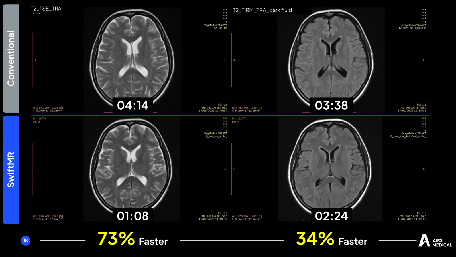
Some sequences could be accelerated much further than the clinic average. A brain T2 axial acquisition fell from 4:14 to 1:08, a 73% reduction, while preserving gray–white matter contrast. The reconstructed FLAIR was 34% faster and appeared less noisy than the original.
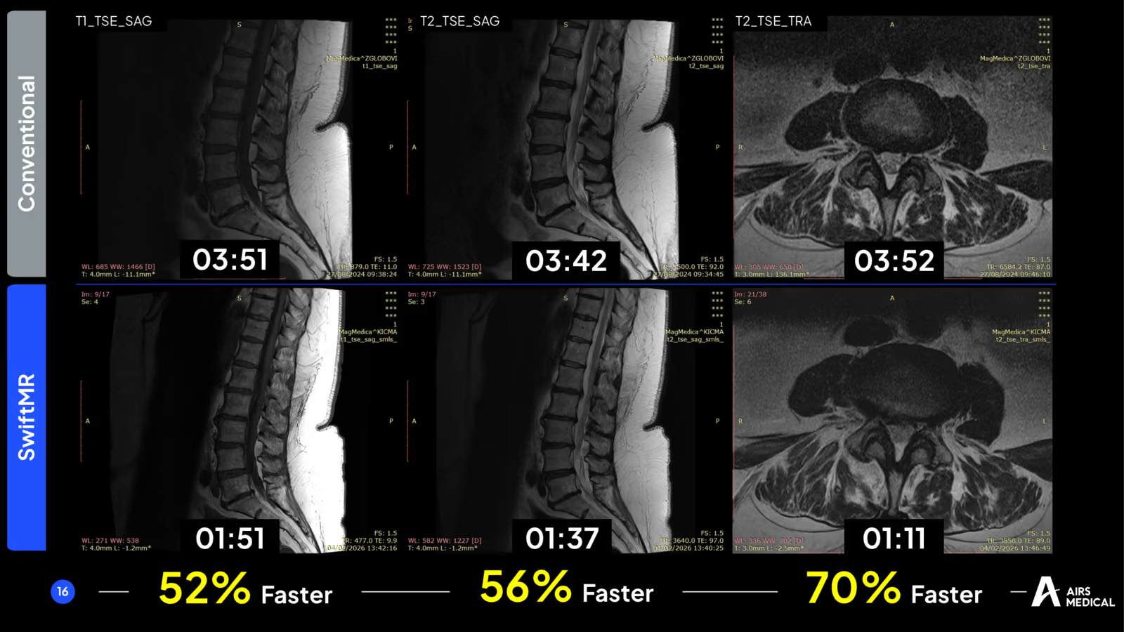
The lumbar spine showed both the opportunity and its limit. The three core sequences were substantially faster, including a 70% reduction on the T2 axial. At that most aggressive setting, however, there was less signal in the dural sac than in the original image.
“The holy trinity of the lumbar spine,” as Dr. Nosek calls the T1 and T2 sagittals and the T2 axial.
SwiftMR let Mag-Medica make that trade-off sequence by sequence rather than accept one setting for the whole protocol. Where speed mattered most, acceleration was pushed to its limit; where maximum signal mattered more, acceleration could be dialed back instead.
When detail mattered, Mag-Medica chose image quality
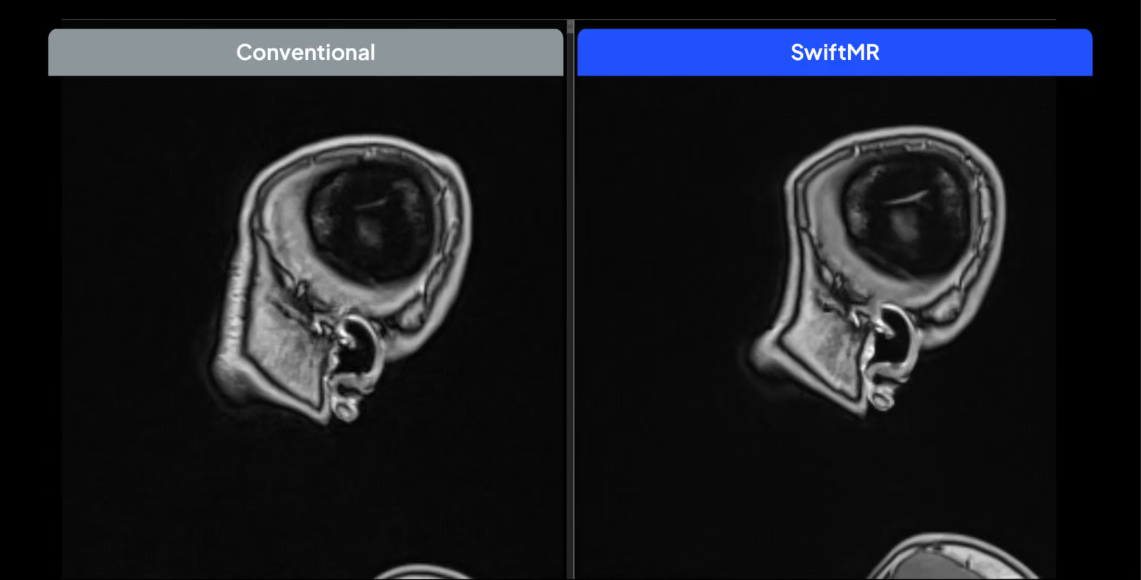
Other examinations called for a different balance. With SWI and T1 MPRAGE, scan-time savings were modest, while the reconstructed images showed less noise and clearer fine structures. In neuro-oncology, that clarity can help in assessing small leptomeningeal metastases.
MR angiography provided one of the clearest examples. The acquisition was 30% faster, yet small lenticulostriate perforating arteries obscured by noise in the original were visible in the reconstructed image. The processed data also produced clearer 3D vascular reconstructions.
Sharper detail where 1.5T often struggles
The same choice carried into body imaging. Small-field-of-view pelvic studies can be challenging at 1.5T. In the prostate case shown at ECR, SwiftMR produced a sharper, less noisy image in which a PI-RADS 5 lesion in the left peripheral zone remained clearly demarcated. “There is absolutely no noise in the images,” Dr. Nosek said of the study, “which is not something you would expect from such an old 1.5 Tesla system.”
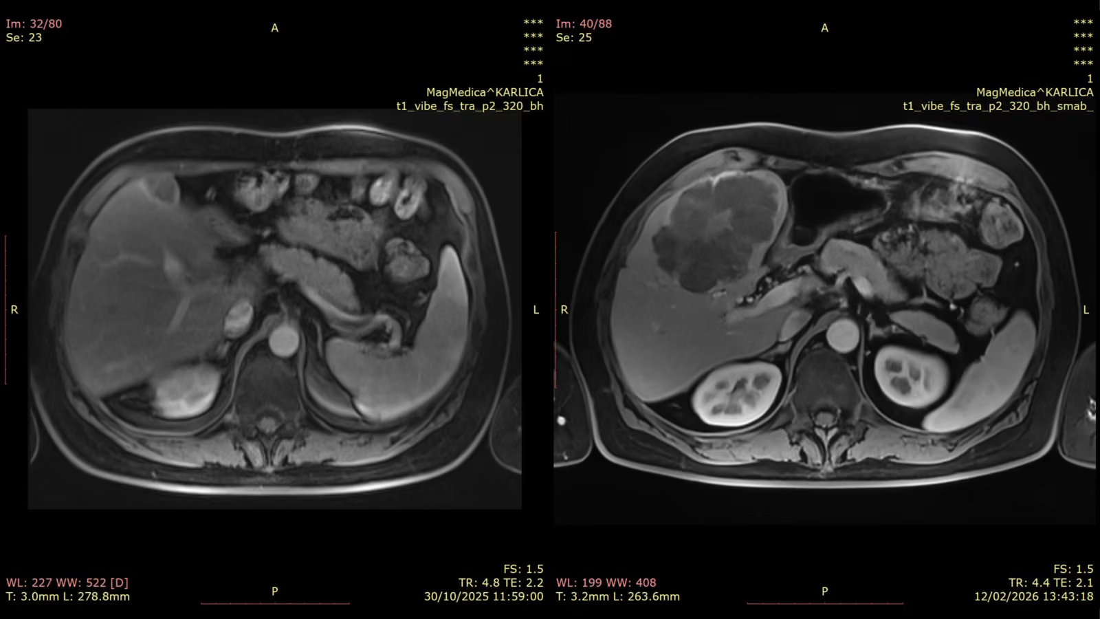
Abdominal oncology created a different constraint. These sequences were already governed by how long the patient could hold their breath, often not very long in an oncology population, so there was little acquisition time to save. The benefit showed up instead as sharper borders around the vessels and pancreas, and far less noise in the visceral fat.
“This is a godsend when it comes to finding these very small peritoneal and omental deposits that we see all the time in oncology.”Dr. Igor Nosek
Abdominal oncology: the two studies are from different follow-up dates and the disease has progressed, so the comparison is in image character rather than lesion size.
More capacity, clearer images, no new scanner
SwiftMR changed more than individual scan times. It changed what Mag-Medica could expect from the Avanto at the center of its practice. On the same scanner, average acquisition time fell by 39.5%, daily capacity rose by about 20%, and the team gained sharper, less noisy images across a wide range of examinations.
Just as importantly, the gain was flexible: speed when the schedule demanded capacity, image quality when fine detail mattered. SwiftMR became part of day-to-day protocol decisions across the practice.
Mag-Medica kept its “trusty workhorse.” What changed was how many patients it could serve, and what its radiologists could ask of the images.
“Your images will be sharper, they will be less noisy, and they will be of better resolution, which will enable you to have more accurate detection of any kind of changes.”Dr. Igor Nosek
See what SwiftMR® could change at your site
Every scanner, protocol and workflow is different. Share your clinical priorities with our team and evaluate SwiftMR on your own images to see where it could improve acquisition time, image quality and capacity in your practice.
Results reflect Mag-Medica’s measurements on a Siemens Magnetom Avanto 1.5T, as presented by Dr. Igor Nosek at the ECR 2026 AI Lightning Talk. Actual results vary by scanner, sequence and protocol.