Liver DCE MRI — Same Scan. More to See.
Arterial phase imaging requires precise timing and sufficient image quality to evaluate enhancement patterns, hepatic vasculature, and lesion conspicuity within a limited acquisition window.
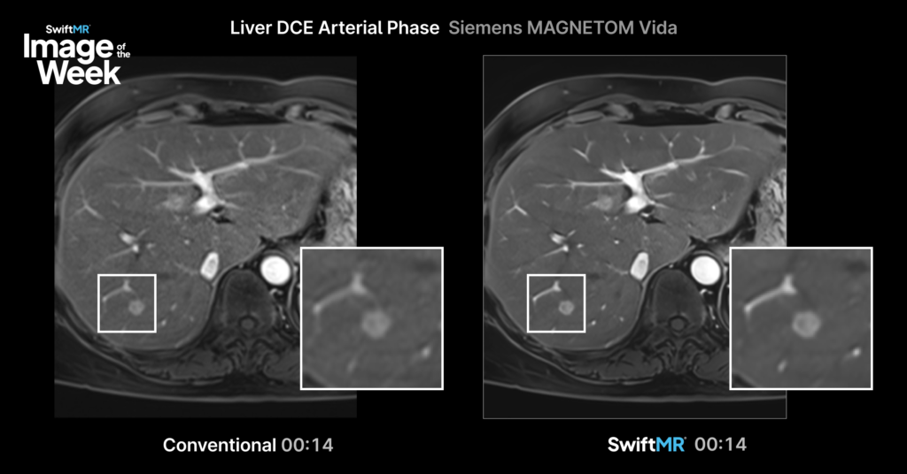
This clinical example compares standard arterial phase liver imaging with a SwiftMR-reconstructed image acquired on a Siemens MAGNETOM Vida 3.0T using a standard 3D VIBE sequence (1.0 × 1.4 × 6.0 mm).
Areas of Interest
This comparison highlights differences in:
- Hepatic vessel delineation
- Background signal within the liver parenchyma
- Overall lesion conspicuity
Because both images were acquired using the same scanner and protocol, this comparison provides an opportunity to assess how image reconstruction may influence visualization of clinically relevant structures during arterial phase imaging.
Clinical Considerations
For radiologists evaluating liver lesions, arterial phase image quality can directly impact visualization of hepatic vessels, lesion characterization, and overall diagnostic confidence.
Because both images were acquired using the same scanner and protocol, this comparison provides an opportunity to assess how SwiftMR reconstruction may influence visualization of clinically relevant structures during arterial phase imaging.
Differences in vessel delineation, background liver signal, and lesion conspicuity can be evaluated without changes to the acquisition protocol, allowing radiologists to assess the potential impact of advanced image reconstruction on routine liver MRI examinations.
Image Details
- Scanner: Siemens MAGNETOM Vida 3.0T
- Sequence: 3D VIBE
- Resolution: 1.0 × 1.4 × 6.0 mm
- Comparison:
- Standard Reconstruction
- SwiftMR Reconstruction
For additional clinical examples, explore the AIRS Medical clinical image gallery or contact Keaur Patel to discuss specific MRI protocols and imaging applications.