Metal Hardware MRI: Same Protocol. Recovering Signal Where It Counts
Metal hardware is one of the biggest image quality challenges in MRI, and one of the least discussed. When a patient has a hip replacement, spinal rods, or orthopedic hardware of any kind, the scan gets harder. The metal distorts the magnetic field around it, creating artifacts that can obscure exactly the anatomy you are trying to see. Technologists work around this with specialized sequences and protocol adjustments, but there is an inherent tradeoff: the techniques that reduce artifacts also reduce signal, and the result is noisier and harder to read confidently.
This compromise impacts patients directly. Critical clinical details — such as hardware stability, peri-implant infection, or spinal healing — often become obscured by artifact and noise. SwiftMR addresses this without changing how your site already scans.
Areas of Interest
This comparison highlights differences in:
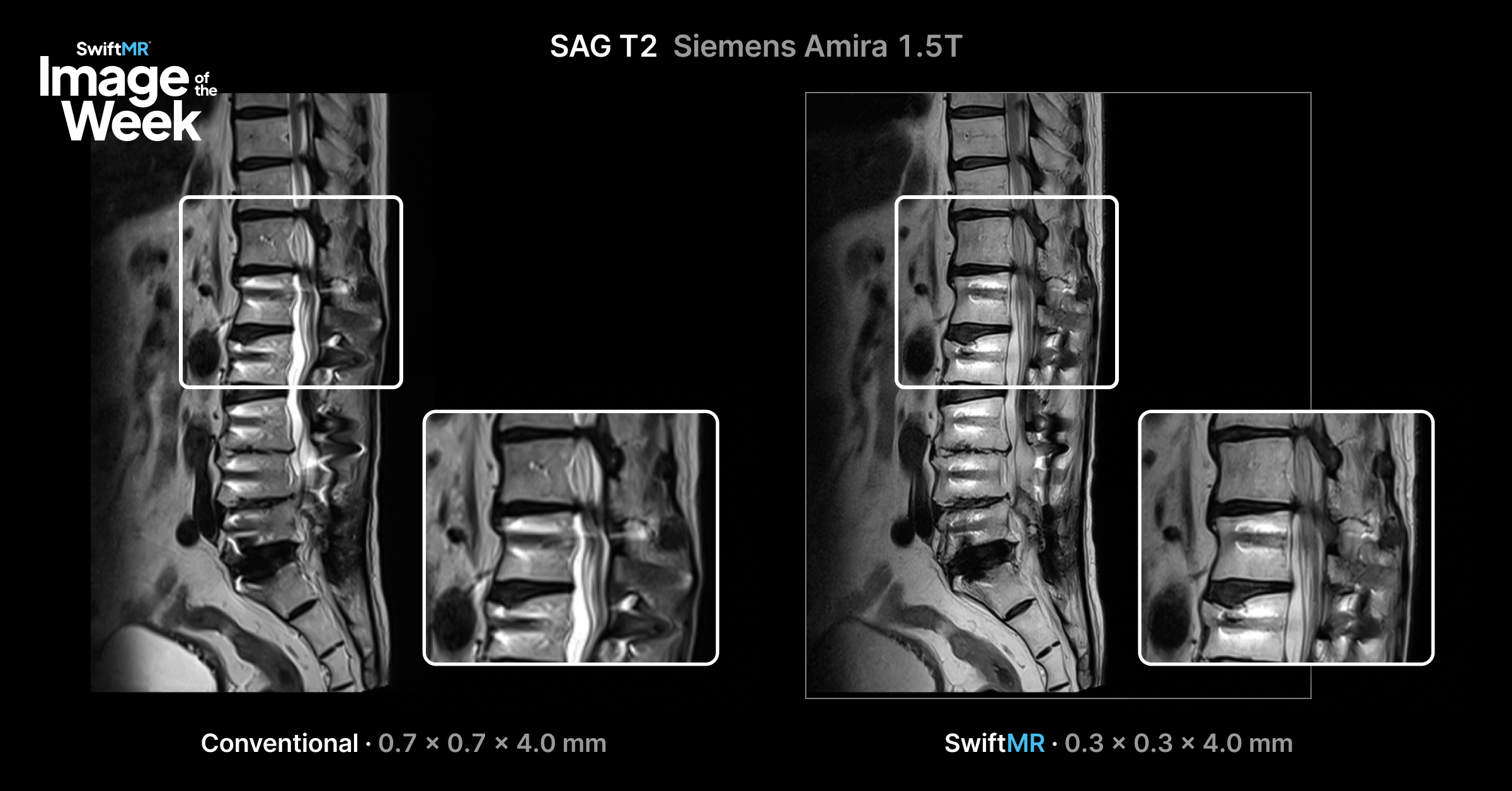
- Signal recovery in high-bandwidth, artifact-reduced acquisitions
- Through-plane resolution and achievable slice thickness near hardware
- Visualization of peri-implant anatomy, cortical margins, and overall detail
Clinical Considerations
SwiftMR is compatible with the metal artifact reduction sequences already available on GE, Siemens, Philips, Canon, and Fujifilm scanners — MARS, SEMAC, MAVRIC, and others. Whatever protocol your technologists use today, SwiftMR works with it. Its reconstruction recovers the SNR that high-bandwidth, artifact-reduction acquisitions cost you, so image quality improves without adding scan time or requiring protocol overhauls.
For through-plane resolution specifically, SwiftMR lets sites scan at thinner slices than would otherwise be practical. Thinner slices mean better anatomical detail near hardware, less partial volume effect, cleaner tissue margins, and more diagnostic confidence. Historically, sites have had to choose between slice thickness and signal. SwiftMR takes that tradeoff off the table.
Because SwiftMR integrates with existing acquisition protocols, the potential impact of advanced reconstruction can be assessed without changes to how the site already scans. For facilities doing volume in joint replacement follow-up, spine surgery, or orthopedic oncology, that is a meaningful improvement in both image quality and throughput, without asking the team to change their workflow.
Image Details
- Compatible sequences: MARS, SEMAC, MAVRIC, and others
- Platforms: GE, Siemens, Philips, Canon, Fujifilm
- Comparison: Standard Reconstruction vs. SwiftMR Reconstruction
For additional clinical examples, explore the AIRS Medical clinical image gallery or contact Keaur Patel to discuss specific MRI protocols and imaging applications.