Solving the variability gap in brain volumetry
Measurements Clinicians Can Trust
Stop scheduling around your scanners
Sample reports
Built on a proven foundation
Track brain health with confidence
Track atrophy across scanners over time
Catch what conventional approaches miss
Deeper analysis beyond whole-brain volumes
Scan anywhere
Even utilization
Better for patients
Conventional methods can lead to a 15–20% difference between scanners. With SwiftSight, achieve consistent brain volumetry and quantification regardless of the manufacturer or field strength.
Hippocampal Volume (ml) across scanners
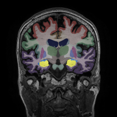
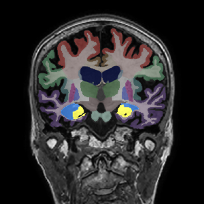
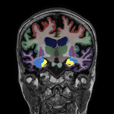
Same patient scanned on three different scanners
SwiftMR delivers sharp images that look the same, regardless of which scanner you used. SwiftSight expands what the MRI reveals, providing consistent, highly-accurate brain health measurements you can rely on for longitudinal tracking.
A female patient aged 72, 76, and 77 was scanned on three different scanners across five years.
Despite the three different vendors, SwiftSight with SwiftMR normalization produced a clean, monotonic atrophy curve — eliminating scanner-induced volume variations that confound longitudinal monitoring.
SwiftSight segments structures that conventional tools cannot reliably quantify — including brainstem, choroid plexus volumetry, and full lobar breakdown for cortical and subcortical regions.
Patients go to whichever scanner is open next — no more waiting for a specific machine.
Spread volume across your entire fleet instead of overloading some scanners while others sit idle.
Shorter waits, fewer reschedules, and scans at whichever location is most convenient.
Patient waits 3 weeks for a specific scanner
Available today, but may yield variation
Available tomorrow, but may yield variation
Patient can scan today on the first available scanner
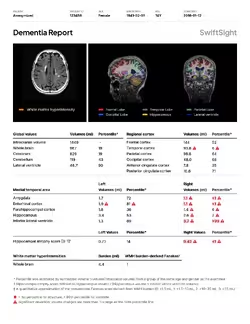
Longitudinal volume tracking with percentile comparisons across brain regions
Regional volume analysis for Alzheimer's and related neurodegenerative conditions
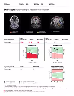
Quantitative assessment of hippocampal asymmetry for detecting lateralized structural abnormalities and supporting neurological diagnosis
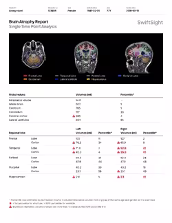
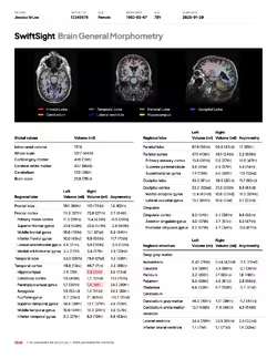
Comprehensive brain morphometry analysis providing global and regional volumetric assessment across brain structures
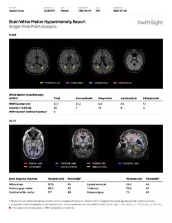
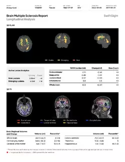
WMH burden quantification and regional distribution analysis
Lesion tracking, volume changes, and longitudinal MS progression monitoring
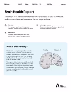
Simplified report designed to help patients understand their own brain health
ARIA Report · DTI · Metabolic · Spine · MSK
SwiftSight is powered by SwiftMR, the most widely deployed vendor-neutral MRI AI platform in the US
“AIRS team provides high quality products with superior customer service and knowledge.”
3T Radiology & Research, Co-founder
“Our team of radiologists and technologists were able to easily partner with AIRS Medical team.”
Invision Sally Jobe, Lead MRI Technologist
“(Thanks to SwiftMR,) I honestly can't believe I'm saying the MRI experience was a 10, but it really was that good.”
East River Medical Imaging, Patient