Smarter MRI, from end to end
Higher efficiency. Sharper images. Deeper insights.
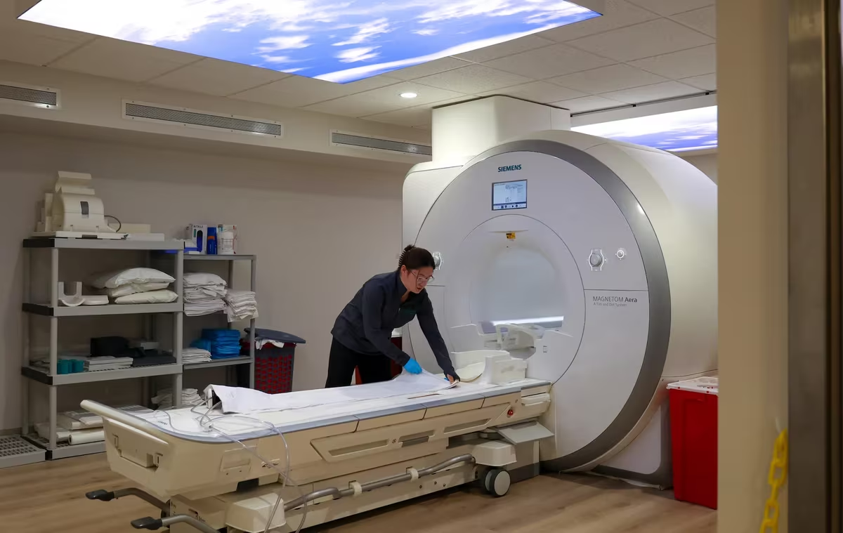
Book a demoMRI is powerful.
But the experience is broken.
For patients, providers, and radiologists — the MRI workflow hasn’t kept up with the promise of the technology.

Scans take too long

Upgrades cost a fortune

Image quality varies dramatically

Patients can’t understand their results
One scan. Four layers of intelligence.
SwiftMR
AI-powered image reconstruction that reduces scan time by up to 50% while improving image quality. Works on any scanner, any vendor.
SwiftSight
Automated volumetric analysis for brain, spine, and metabolic health. Percentile mapping, longitudinal tracking, and cross-scanner consistency.
SwiftRead
AI-translated reports that turn complex radiology findings into plain language patients can understand and act on.
Patient Portal
A web portal where patients access reports, explore findings, track health over time, and connect with your center.
CONVENTIONAL
04:53
SWIFTMR
01:46
SCAN TIME
64% FASTER
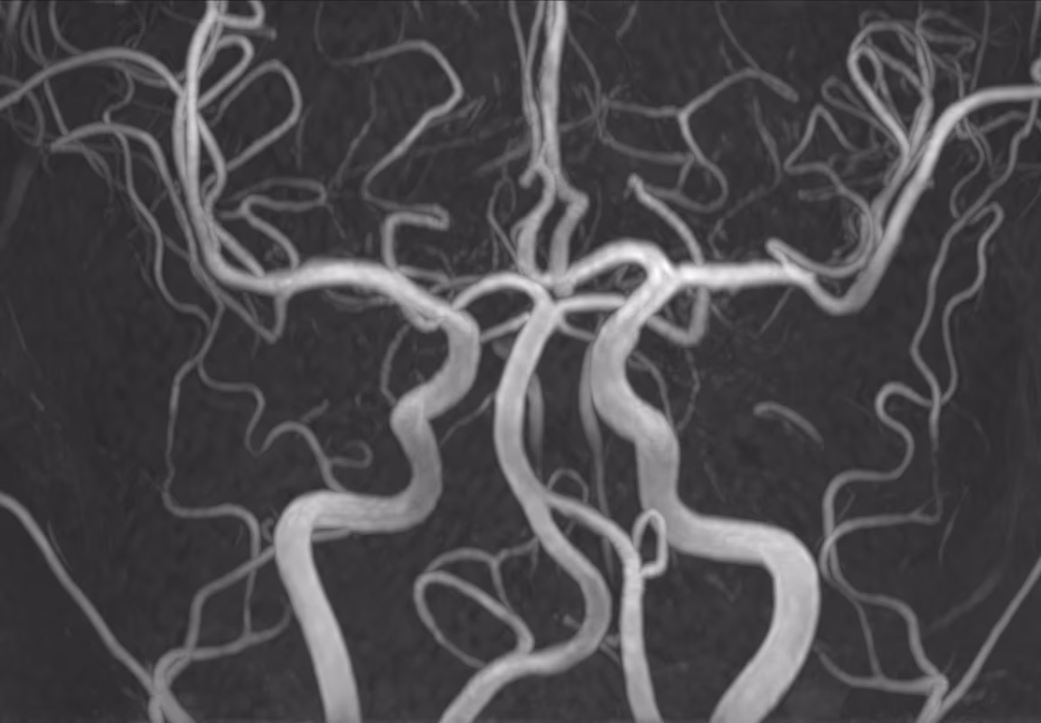
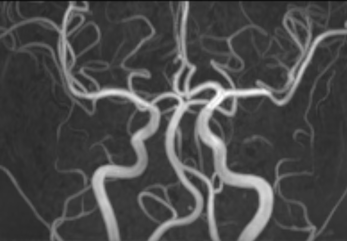
Brain MRA, 3.0T Siemens MAGNETOM Skyra
3D TOF, (L) 0.6×0.8×1.2 mm, (R) 0.6×0.8×1.2 mm, MIP
Cut scan times in half. Improve image quality.
SwiftMR uses deep learning to reconstruct MRI images with less noise and higher resolution. It works on every sequence, every body part, every vendor.
See image gallery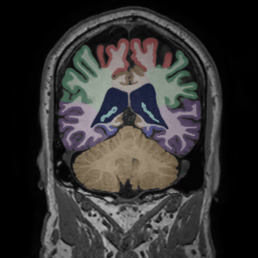
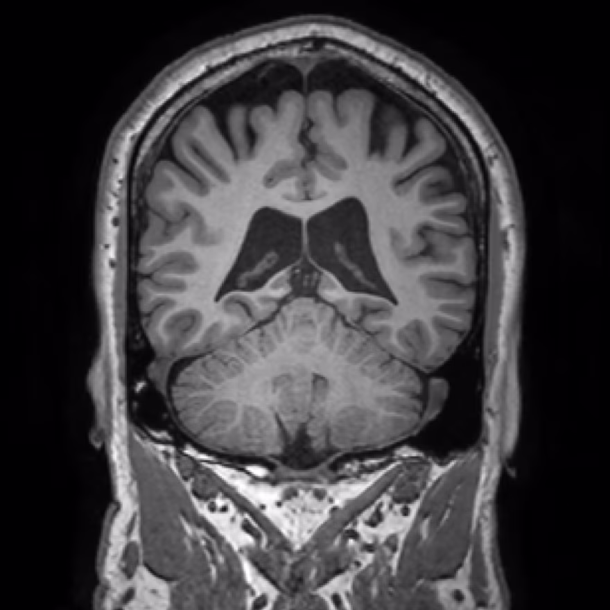
INPUT
SWIFTSIGHT
SEGMENTATION
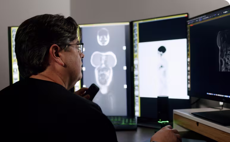
Quantitative imaging clinicians can rely on
SwiftSight delivers brain volumetry, body composition analysis, and more — with cross-scanner consistency that existing solutions can’t match. When cross-scanner variation goes from 16% to less than 2%, reliable longitudinal tracking becomes possible.
Learn about SwiftSightCLINICAL HISTORY: Chronic headaches, r/o intracranial pathology. Hx of mild TBI 2019. Pt reports intermittent visual disturbances x 3 months.
TECHNIQUE: Multiplanar multisequence MR imaging of the brain was performed without IV contrast on a 3T Siemens Magnetom Prisma scanner. Sequences include sagittal T1 MPRAGE, axial T2, axial FLAIR, axial DWI/ADC, axial SWI, coronal T2.
FINDINGS:
No evidence of acute infarction on diffusion-weighted imaging. No intra- or extra-axial hemorrhage identified on SWI sequences. Brain parenchyma demonstrates normal signal intensity on all sequences. Gray-white matter differentiation is preserved. No focal parenchymal signal abnormality. No mass effect or midline shift. Ventricular system is normal in size and morphology for patient age. No evidence of obstructive or communicating hydrocephalus.
Basal cisterns are patent. Cerebellar tonsils are in normal position relative to the foramen magnum. Corpus callosum is intact with normal morphology. Pituitary gland measures within normal limits, no focal lesion. Major intracranial flow voids are preserved bilaterally. Calvarium and skull base demonstrate no osseous abnormality.
Visualized paranasal sinuses demonstrate mild mucosal thickening in bilateral maxillary sinuses. Mastoid air cells are clear. Orbits are grossly unremarkable to the extent visualized.
IMPRESSION:
1. No acute intracranial abnormality. No evidence of hemorrhage, infarction, or mass lesion.
2. No significant signal abnormality identified within the brain parenchyma.
3. Incidental mild maxillary sinus mucosal thickening, likely inflammatory/allergic in etiology. Clinical correlation recommended.

Reports patients can actually understand
SwiftRead translates radiology reports into patient-friendly language with body maps, color-coded findings, and clear next steps. Improve your patients’ satisfaction and enable them to take better control of their health.
Request informationReal outcomes. Proven results.
Extending the life of their scanners instead of buying new
“ISJ uses a multi-year capital equipment prioritization process, and needed a solution that would help us use our existing equipment to quickly meet rapid increases in demand.”
Geoff Lawton, CEO
Additional $102K new revenue in the first month
“The return on investment has been fantastic. We’ve increased our margins significantly with less hours and more scans.”
Humberto Carrion
Co-founder
13-hour days down to 10 with the same volume
“Our staff finally has a work-life balance again. We can see more patients and still leave on time.”
Kevin Taylor
TaylorMED MRI, Owner
Trusted by radiologists & operators
“We were able to accomplish all of the goals within the first two months — increasing throughput, reducing business hours and backlogs, and improving the patient’s overall experience.”
Humberto Carrion
Co-founder, 3T Radiology & Research
“AIRS Medical helped us achieve dual objectives — improving efficiency while conserving capital. We’re operating more seamlessly on the same devices.”
Geoff Lawton
CEO, Invision Sally Jobe
“I am absolutely blown away every time I use SwiftMR! This is a game changer, and it instantly brought us up to the next level of quality and productivity.”
Allen Long
Lead MRI Tech, Trinity Health
“I honestly can’t believe I’m saying the MRI experience was a 10, but it really was that good.”
Michael
Patient, East River Medical Imaging
“I didn’t think it was possible to increase throughput without compromising image quality, but it is.”
Chris Beaulieu
Operations, Naugatuck Valley Radiology