KCR (2023)
PURPOSE
This study evaluates the effects of deep learning (DL)-based image reconstruction on the reproducibility and predictive capabilities of radiomic features derived from knee MR images. For evaluation, two different tasks, age prediction and disease classification were performed by comparing radiomic features from standard-of-care (SOC) and deep learning (DL)-processed images.
MATERIALS AND METHOD
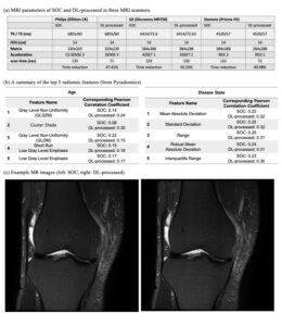
This analysis utilized de-identified data, waiving the requirement for IRB approval. 45 patients underwent knee MRI exams on three different 3T MR scanners, both with SOC and DL-accelerated protocols. Coronal T2 fat-saturated turbo spin-echo (TSE) knee MR images were acquired, with the DL-accelerated protocol achieving a 41% scan time reduction. These accelerated scans were then processed using DL-based MR reconstruction software (SwiftMR, AIRS Medical) that functions image denoising and super-resolution.
We extracted a total of 94 radiomic features using Pyradiomics package and aimed to predict two target parameters: age and disease state. The patients’ age and disease state in the bone marrow region were labeled from a radiologist’s examination.
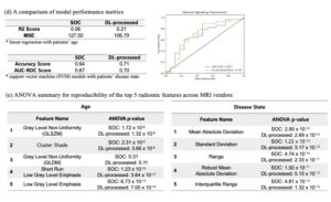
For each target parameter, we selected the top five radiomic features based on the sum of Pearson correlation coefficients from SOC and DL images. Linear regression was applied for age prediction and evaluated through R2 scores and mean squared errors (MSE), while a support vector machine (SVM) model was employed for disease classification, assessed by accuracy and AUC-ROC. Addressing radiomics’ inherent reproducibility issue, we grouped subjects by the MRI scanner vendor and performed ANOVA on the top five radiomic features across these groups.
RESULTS
DL-processed images surpassed SOC images in age prediction with higher correlations and lower MSE (R2 score: 0.21 vs. 0.06, MSE: 106.79 vs. 127.50), and in disease classification with superior SVM model performance (accuracy: 0.71 vs. 0.64, AUC-ROC: 0.70 vs. 0.67). Notably, reproducibility analysis revealed DL-processed images provided improved consistency across varying scanner vendors, as indicated by lower ANOVA p-values.
CONCLUSION
DL-based image improvement of knee MRIs can enhance the identification of cartilage lesions, without affecting overall diagnostic performances as correlated with arthroscopic results.