AIRS Medical at RSNA 2024
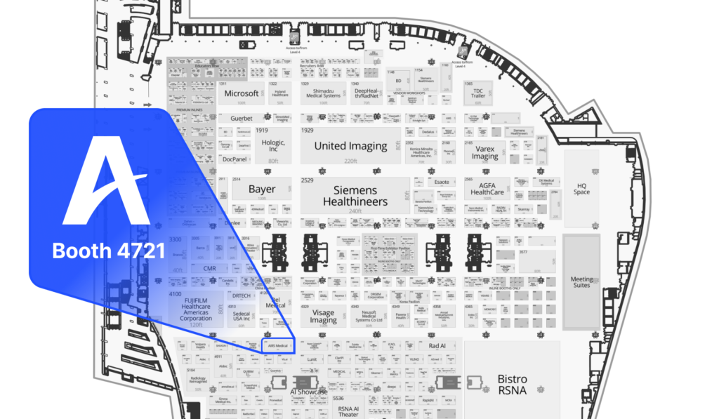
Booth #4721 (South Hall)
Explore our latest innovations in medical imaging and discover how the award-winning SwiftMR™ can transform your radiology practice.

Week at a Glance
Sunday
December 1
Monday
December 2
Tuesday
December 3
Wednesday
December 4
Thursday
December 5
Sunday
December 1
Lunch & Learn
11:45am – 12:45pm CT
South Building, Room S403A
SwiftMR™: How a Simple Software Upgrade Can Improve MRI Image Quality, Boost Revenue, and Enhance the Patient Experience
This session is ideal for radiologists, operations leaders, IT specialists, and healthcare administrators looking for ways to stay ahead in the hyper-competitive medical imaging field.
Amish Doshi, MD (Moderator)
Chief of the Division of Neuroradiology at Mount Sinai
Timothy Deyer, MD
Chief Medical Information Officer at East River Medical Imaging
Chris Beaulieu
Operations Director at Naugatuck Valley Radiological Associates
Kevin Yang, PhD
VP, Head of Clinical Research
AI Theater Presentation
3:00pm – 3:15pm CT
Advancing Neuroradiology: Clinical Research and Validation of SwiftMR in a University Hospital
Dr. Choi will detail his experiences utilizing SwiftMR™ for various neuroimaging applications in a University Hospital setting and will share real-world research and validation efforts surrounding SwiftMR’s AI algorithm.
Seung-Hong Choi, MD, PhD
Professor in the Department of Radiology at Seoul National University
Monday
December 2
AI Theater Presentation
11:00am – 11:15am CT
Over $200,000 in Additional Revenue: How a Leading Diagnostic Imaging Center Revamped its Practice in Two Months with SwiftMR™
This session is built for operations leaders and decision makers at radiology practices seeking creative ways to generate additional revenue.
Daryl Eber, MD
Co-founder of 3T Radiology & Research
Tuesday
December 3
Poster Presentation (#9473)
12:45pm – 1:15pm
Enhancing Bone MRI: Advanced Edge Detection, CS/CAIPI VIBE Sequences, and Deep Learning Reconstruction Across Diverse Models.
Enhancing Bone MRI: Advanced Edge Detection, CS/CAIPI VIBE Sequences, and Deep Learning Reconstruction Across Diverse Models
Authors: Yoon YE, et al.
This work focused on exploring the feasibility of using 3D MR imaging of the shoulder combined with SwiftMR application for high resolution imaging of the bone. Computed tomography (CT) is the standard choice for imaging small bone lesions of the shoulder due to its superiority in visualizing electron density and in-plane resolution. However, since CT exposes patients to ionizing radiation, it may be clinically advantageous if MR could overcome the inherent low signal-to-noise ratio observed during bone imaging dur to low proton density and short T2 decay times. SwiftMR was applied retrospectively to 3D MR images of the shoulder, and the results have shown that the image quality and diagnostic features become comparable to that of the CT.
Thursday
December 5
Poster Presentation (#4855)
12:15pm – 12:45pm
All-in-one Deep Learning Framework for MR Image Reconstruction.
All-in-one Deep Learning Framework for MR Image Reconstruction
Authors: Geunu Jeong, Hyeonsoo Kim, Joonyoung Yang, Kyungeun Jang, Jeewook Kim (AIRS Medical)
This research introduces a novel, all-in-one deep learning framework for MR image reconstruction, enabling a single model to enhance image quality across multiple aspects of k-space sampling and to be effective across a wide range of clinical and technical scenarios. Multi-dimensional degradation was applied to raw k-space data to generate training input. This process involved a combined application of noise addition and multiple patterns of undersampling (uniform, random, kmax, partial Fourier, elliptical), with each method being applied across a range of factors to cover extensive sampling scenarios. Contextual data, including scan parameter information, were prepared to serve as auxiliary input to address the challenges posed by the unique learning task for each training pair, which arise from the varied degradation scenarios. The expected noise reduction factor for each training pair was mathematically derived and used as additional contextual data for tunable denoising. The U-Net was modified to include an additional pathway for integrating contextual data. The proposed model enhances image quality in a multi-dimensional manner and is compatible with a broad spectrum of scenarios, including various vendors, pulse sequences, scan parameters, and anatomical regions.
Oral Presentation (#14714)
1:30pm – 2:30pm
Optimizing 3d t1 mprage processing for robust differentiation of alzheimer’s disease from cognitively normal in older adults: the impact of advanced deep learning-based reconstruction and segmentation.
Optimizing 3d t1 mprage processing for robust differentiation of alzheimer’s disease from cognitively normal in older adults: the impact of advanced deep learning-based reconstruction and segmentation
Authors: Seoyoung Lim, Woojin Jung, Seoyeon Park, Kyungtae Lee, Geunu Jeong (AIRS Medical), Koung Mi Kang (Seoul National University Hospital)
This study aimed to enhance the differentiation between Alzheimer’s disease (AD) and cognitively normal (CN) older adults using deep learning (DL)-based algorithms applied to MRI. Utilizing the OASIS-2 open dataset, which included 3D T1-weighted MRI scans from 150 subjects aged 60-96 across 373 MRI sessions, we applied DL-based image reconstruction (DLR) and volumetry (DLV) techniques. The results showed a significant performance improvement, with AUC-ROC increasing from 96.8% to 97.5% with FastSurfer, and from 98.1% to 99.0% with DLV after applying DLR. Significantly, the DLR + DLV framework consistently identified the hippocampus and amygdala as the top two significant regions of interest (ROIs) for AD detection, with high reproducibility.
Meet the Experts
Hear directly from leaders in radiology who have successfully integrated SwiftMR™ at their practices.
Discover how they’ve reduced scan times, boosted efficiency, and improved patient outcomes with AI.

East River Medical Imaging
Timothy Deyer, MD, is a board certified Musculoskeletal and Interventional Radiologist specializing in diagnostic musculoskeletal MRI and ultrasound. He serves as Chief Medical Information Officer and Head of Musculoskeletal Interventional Services at East River Medical Imaging, as well as Clinical Assistant Professor of Radiology at Weill Cornell Medical College, Cornell University.

Naugatuck Valley Radiology
Chris Beaulieu is a nationally registered Nuclear Medicine and Radiologic Technologist with ARRT and NMTCB. With over 30 years of experience in clinical and outpatient imaging, he oversees operations at Naugatuck Valley Radiological Associates (NVRA) and serves as a member of the Advisory Board to Naugatuck Valley Community College’s Radiology Program.

3T Radiology & Research
Dr. Eber is Founding Radiologist at 3T Radiology & Research, a leading diagnostic imaging facility in Florida. Board certified in Diagnostic Radiology and Nuclear Medicine, Dr. Eber is a respected leader in his field. Dr. Eber has served as President of the Florida Radiological Society and is an appointed Councilor of the American College of Radiology.

Seoul National University Hospital
Seung-Hong Choi, MD, is Professor of Neuroradiology at Seoul National University. Dr. Choi has made significant contributions to the field of neuroradiology, particularly in the application of cutting-edge imaging technologies. His research interests include brain tumor imaging, neurodegenerative diseases, and developing innovative imaging protocols to enhance diagnostic accuracy.
Where to Find Us


